Copyright
©The Author(s) 2021.
World J Clin Cases. Jun 16, 2021; 9(17): 4453-4459
Published online Jun 16, 2021. doi: 10.12998/wjcc.v9.i17.4453
Published online Jun 16, 2021. doi: 10.12998/wjcc.v9.i17.4453
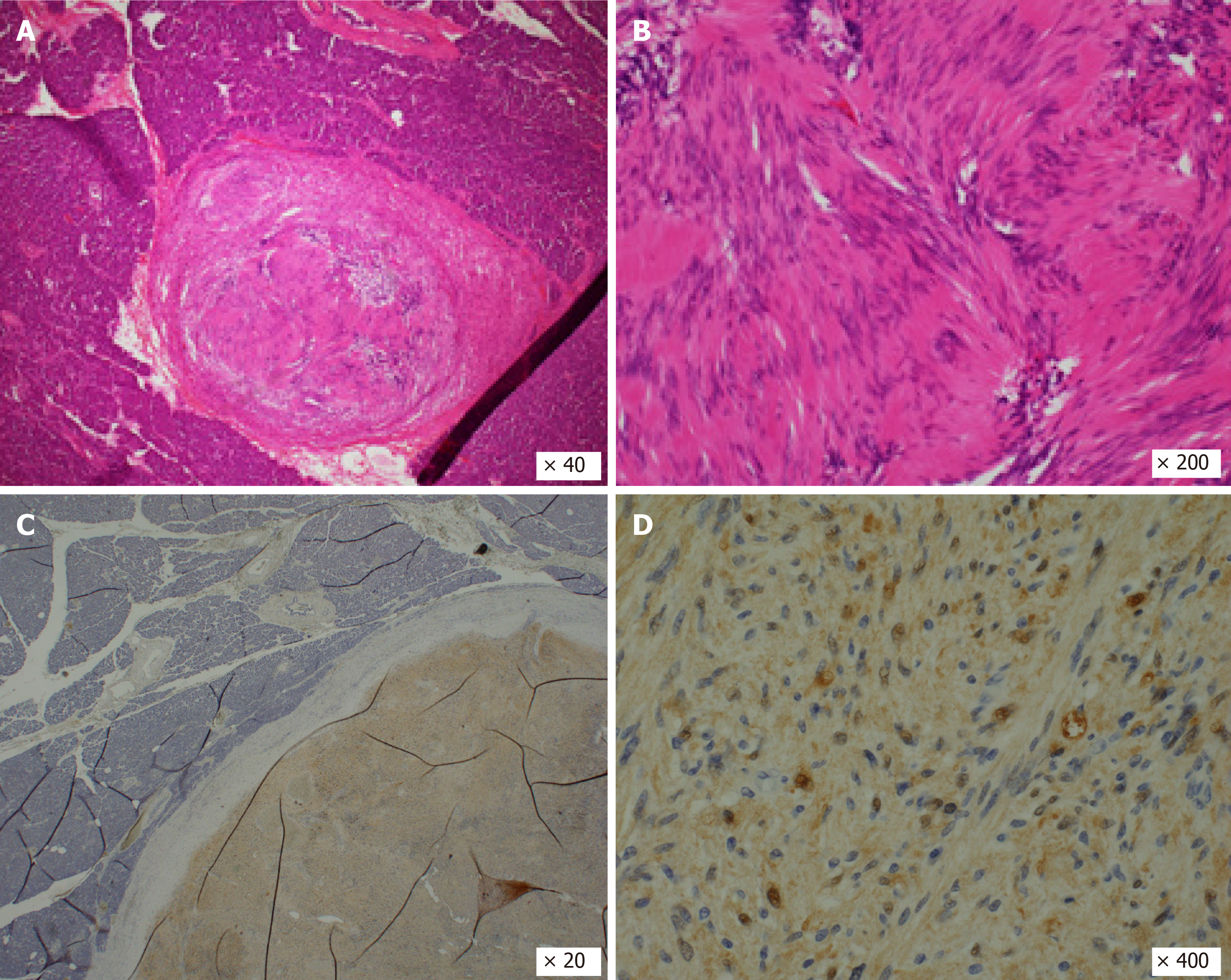
Figure 5 Microscopic histopathological findings.
A and B: Hematoxylin and eosin staining showed a proliferation of spindle-shaped cells in a vague fascicular and haphazard pattern, with palisading arrangement; C and D: Immunohistochemical staining of S100 was positive.
- Citation: Kimura K, Adachi E, Toyohara A, Omori S, Ezaki K, Ihara R, Higashi T, Ohgaki K, Ito S, Maehara SI, Nakamura T, Fushimi F, Maehara Y. Schwannoma mimicking pancreatic carcinoma: A case report. World J Clin Cases 2021; 9(17): 4453-4459
- URL: https://www.wjgnet.com/2307-8960/full/v9/i17/4453.htm
- DOI: https://dx.doi.org/10.12998/wjcc.v9.i17.4453


