Copyright
©The Author(s) 2021.
World J Clin Cases. Jun 16, 2021; 9(17): 4285-4293
Published online Jun 16, 2021. doi: 10.12998/wjcc.v9.i17.4285
Published online Jun 16, 2021. doi: 10.12998/wjcc.v9.i17.4285
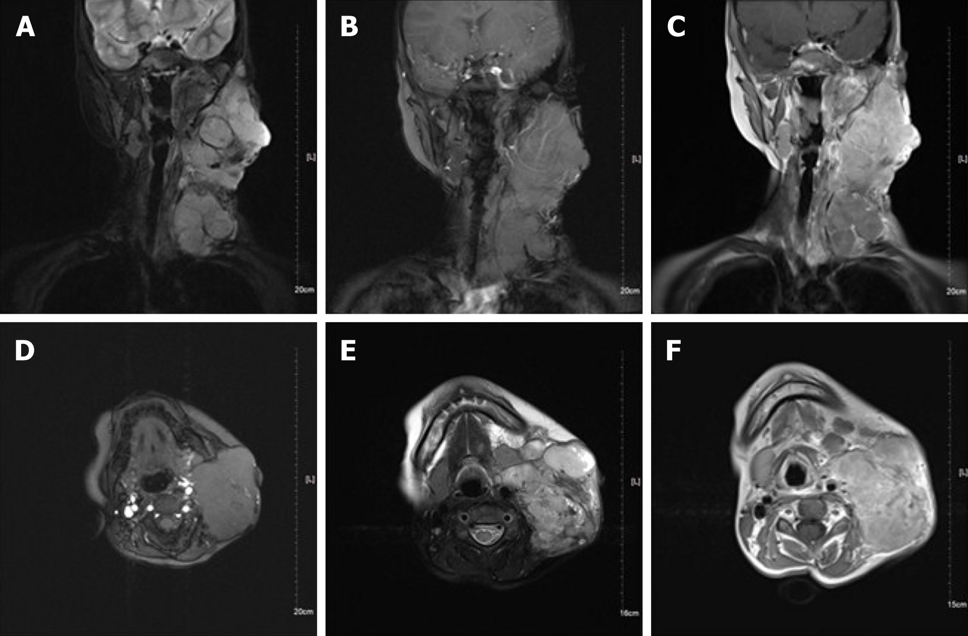
Figure 2 Neck magnetic resonance imaging.
A and D: Magnetic resonance imaging showed many round-like soft tissue masses of different sizes, with the largest measuring 6.5 cm × 5.9 cm × 8.1 cm, in the left parapharyngeal space, carotid sheath area, submandibular region, cervical root, supraclavicular fossa, and supraclavicular region. Some of the lesions fused into masses and the boundary was unclear, with low signal intensity on T1 weighted imaging; B and E: Round-like soft tissue masses showed high signal intensity on fat-suppressed T2 weighted imaging; C and F: The soft tissue mass showed high signal intensity on T2 weighted imaging.
- Citation: Wu GJ, Li BB, Zhu RL, Yang CJ, Chen WY. Rosai-Dorfman disease with lung involvement in a 10-year-old patient: A case report. World J Clin Cases 2021; 9(17): 4285-4293
- URL: https://www.wjgnet.com/2307-8960/full/v9/i17/4285.htm
- DOI: https://dx.doi.org/10.12998/wjcc.v9.i17.4285


