Copyright
©The Author(s) 2021.
World J Clin Cases. Jun 16, 2021; 9(17): 4262-4267
Published online Jun 16, 2021. doi: 10.12998/wjcc.v9.i17.4262
Published online Jun 16, 2021. doi: 10.12998/wjcc.v9.i17.4262
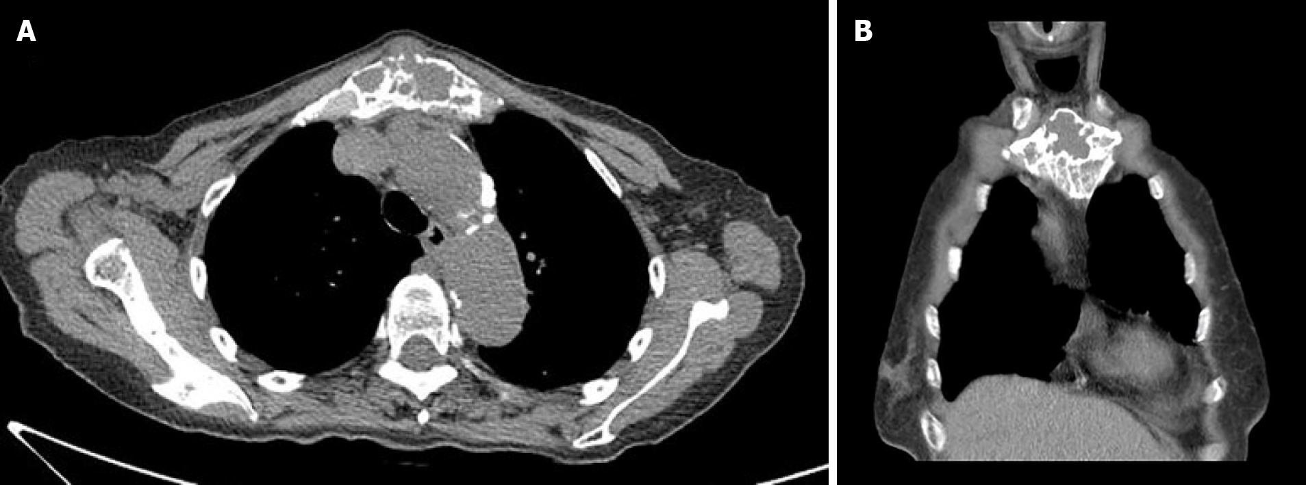
Figure 1 Chest imaging.
A: Axial; B: Coronal thoracic computed tomographic scans showing an expansile and osteolytic mass in the manubrium with cortical destruction.
- Citation: Lin TT, Hsu HH, Lee SC, Peng YJ, Ko KH. Dynamic magnetic resonance imaging features of cavernous hemangioma in the manubrium: A case report. World J Clin Cases 2021; 9(17): 4262-4267
- URL: https://www.wjgnet.com/2307-8960/full/v9/i17/4262.htm
- DOI: https://dx.doi.org/10.12998/wjcc.v9.i17.4262


