Copyright
©The Author(s) 2021.
World J Clin Cases. Jun 16, 2021; 9(17): 4230-4237
Published online Jun 16, 2021. doi: 10.12998/wjcc.v9.i17.4230
Published online Jun 16, 2021. doi: 10.12998/wjcc.v9.i17.4230
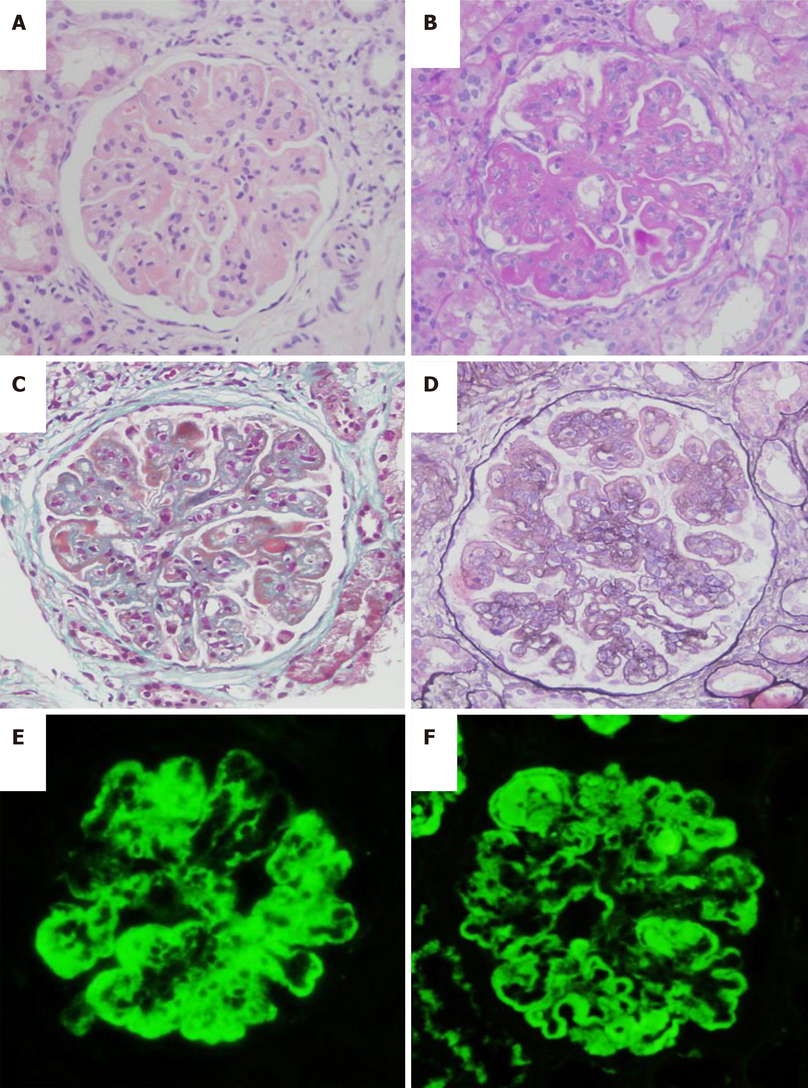
Figure 1 Biopsy pathology.
A: Thickened basement membrane (hematoxylin-eosin staining, × 400); B: Proliferation of mesangial cells and endothelial cells (periodic acid–Schiff stain, × 400); C: Segmental wire loop (Masson, × 400); D: Segmental dual track formation (periodic acid-silver methenamine, × 400); E and F: Granular deposition along the mesangial region and capillary wall (immunofluorescence, × 400).
- Citation: Zhou XS, Lu YY, Gao YF, Shao W, Yao J. Bone marrow inhibition induced by azathioprine in a patient without mutation in the thiopurine S-methyltransferase pathogenic site: A case report. World J Clin Cases 2021; 9(17): 4230-4237
- URL: https://www.wjgnet.com/2307-8960/full/v9/i17/4230.htm
- DOI: https://dx.doi.org/10.12998/wjcc.v9.i17.4230


