Copyright
©The Author(s) 2021.
World J Clin Cases. Jun 16, 2021; 9(17): 4221-4229
Published online Jun 16, 2021. doi: 10.12998/wjcc.v9.i17.4221
Published online Jun 16, 2021. doi: 10.12998/wjcc.v9.i17.4221
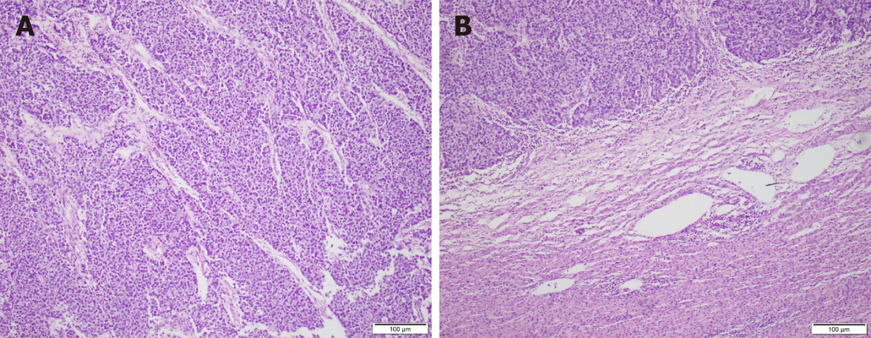
Figure 3 Microscopic images.
A: Poorly differentiated gastric carcinoma (hepatoid adenocarcinoma with neuroendocrine differentiation) with liver metastases; B: Tumor tissue and adjacent liver tissue.
- Citation: Wang H, Zhang CC, Ou YJ, Zhang LD. Ex vivo liver resection followed by autotransplantation in radical resection of gastric cancer liver metastases: A case report. World J Clin Cases 2021; 9(17): 4221-4229
- URL: https://www.wjgnet.com/2307-8960/full/v9/i17/4221.htm
- DOI: https://dx.doi.org/10.12998/wjcc.v9.i17.4221


