Copyright
©The Author(s) 2021.
World J Clin Cases. Jun 16, 2021; 9(17): 4221-4229
Published online Jun 16, 2021. doi: 10.12998/wjcc.v9.i17.4221
Published online Jun 16, 2021. doi: 10.12998/wjcc.v9.i17.4221
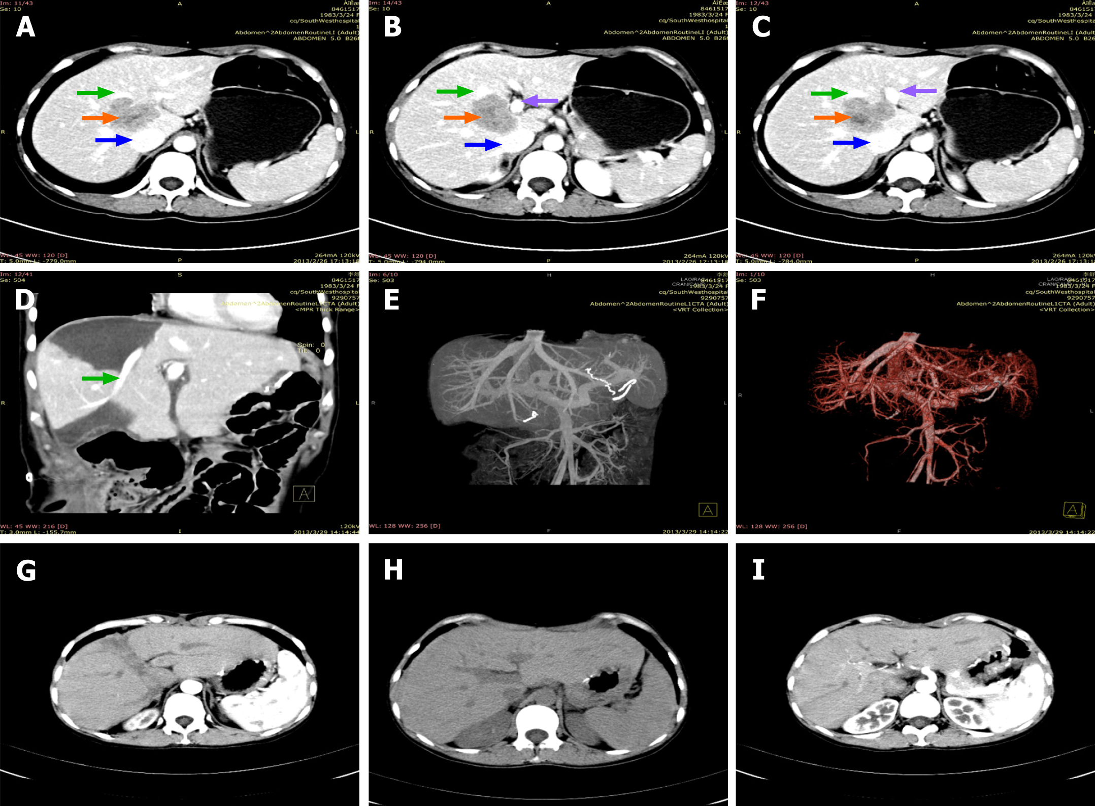
Figure 1 Abdominal computed tomography and magnetic resonance imaging examinations.
A-C: The tumor (pointed by the orange arrow) was located between the first and second hila and it was adjacent to the middle hepatic vein (pointed by the green arrow), inferior R vena cava (pointed by the blue arrow), and the right branch of portal vein (pointed by the purple arrow); D-F: Postoperative imaging examination and upper abdominal angiography showed that the left hepatic vein, middle hepatic vein, right hepatic vein, inferior R vena cava, hepatic artery, and portal vein were intact and blood flow was unobstructed; G-I: Computed tomography results in the first, second, and third year after operation showed that there were no obvious signs of tumor recurrence.
- Citation: Wang H, Zhang CC, Ou YJ, Zhang LD. Ex vivo liver resection followed by autotransplantation in radical resection of gastric cancer liver metastases: A case report. World J Clin Cases 2021; 9(17): 4221-4229
- URL: https://www.wjgnet.com/2307-8960/full/v9/i17/4221.htm
- DOI: https://dx.doi.org/10.12998/wjcc.v9.i17.4221


