Copyright
©The Author(s) 2021.
World J Clin Cases. Jun 6, 2021; 9(16): 4104-4109
Published online Jun 6, 2021. doi: 10.12998/wjcc.v9.i16.4104
Published online Jun 6, 2021. doi: 10.12998/wjcc.v9.i16.4104
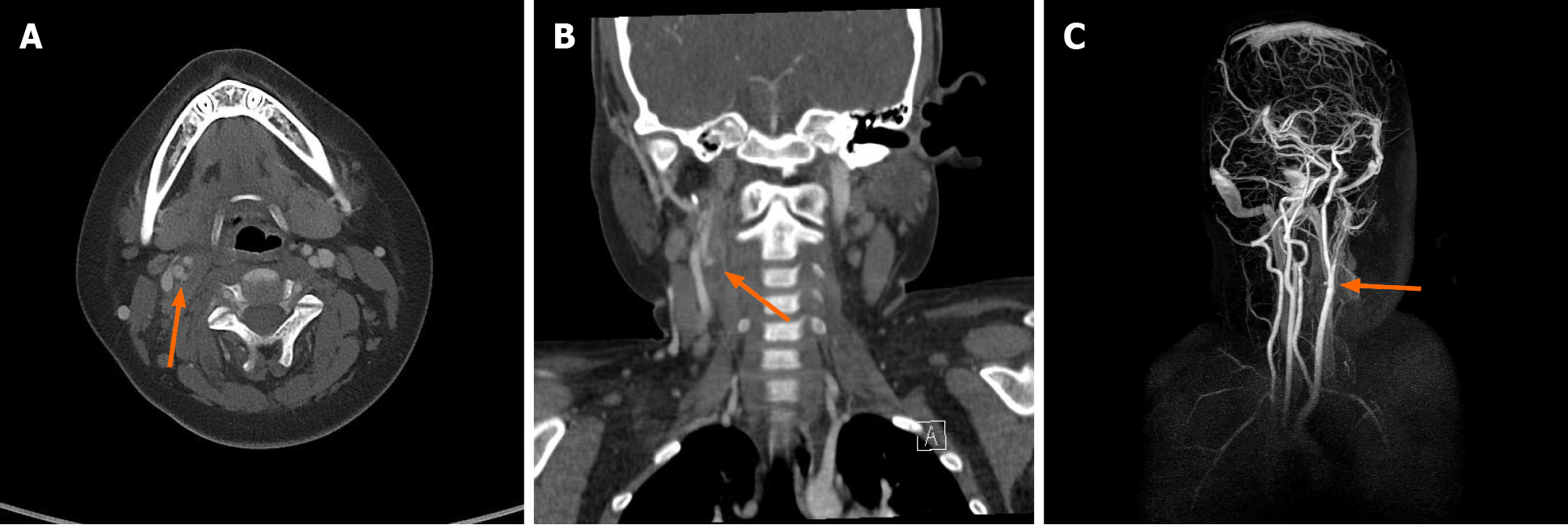
Figure 1 Computed tomography scan and the magnetic resonance angiography of the neck obtained on the second visit of hospital.
A and B: Transverse (A) and coronal section (B) showing the hematoma with pseudoaneurysmal formation of right internal carotid artery (arrow); C: Reconstruction image also showing the pesudoaneurysmal changed right internal carotid artery (arrow).
- Citation: Chung BH, Lee MR, Yang JD, Yu HC, Hong YT, Hwang HP. Delayed pseudoaneurysm formation of the carotid artery following the oral cavity injury in a child: A case report. World J Clin Cases 2021; 9(16): 4104-4109
- URL: https://www.wjgnet.com/2307-8960/full/v9/i16/4104.htm
- DOI: https://dx.doi.org/10.12998/wjcc.v9.i16.4104


