Copyright
©The Author(s) 2021.
World J Clin Cases. Jun 6, 2021; 9(16): 4095-4103
Published online Jun 6, 2021. doi: 10.12998/wjcc.v9.i16.4095
Published online Jun 6, 2021. doi: 10.12998/wjcc.v9.i16.4095
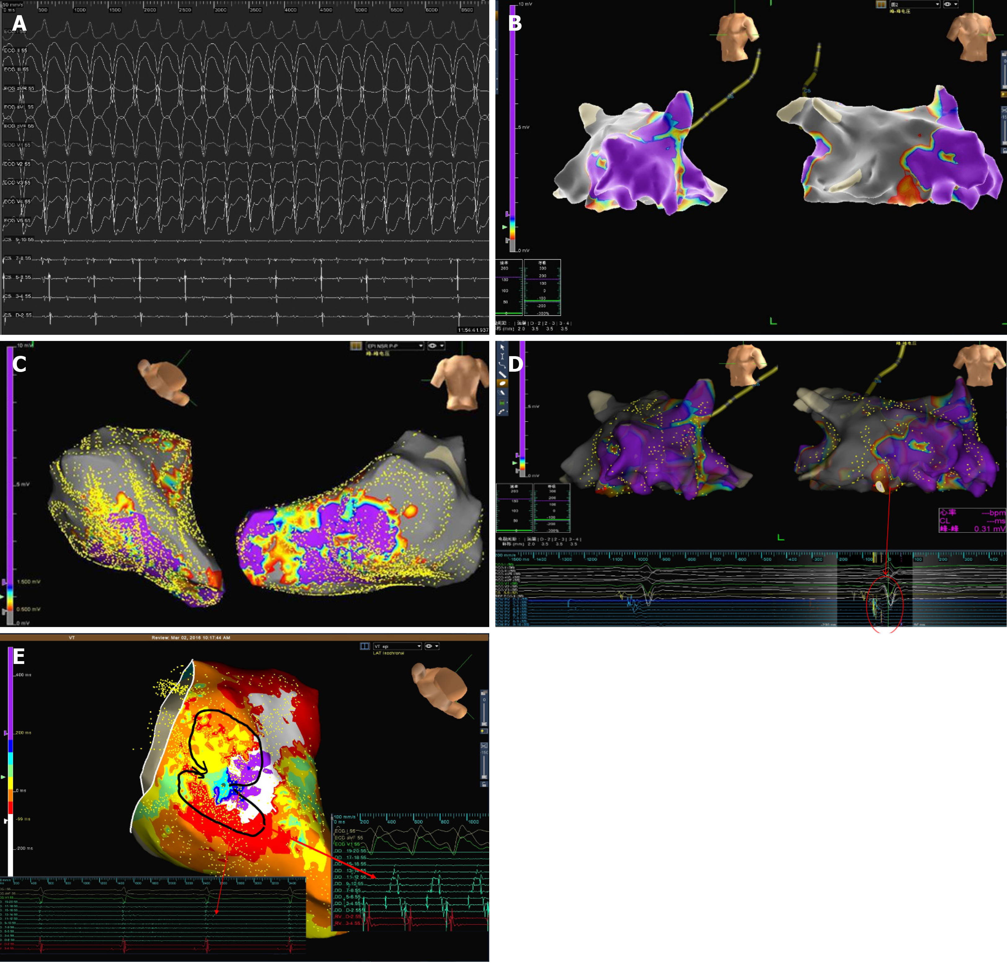
Figure 4 Electrophysiological study results.
A: Multiple inducible right ventricular tachycardias of a focal mechanism; B and C: Endocardial (B) and epicardial (C) 3D-electroanatomic voltage mapping demonstrated scar tissue in the anterior wall, free wall and posterior wall of the right ventricle (gray area); D: 3D electroanatomic voltage mapping showed late potentials (red arrow); E: The focal mechanism of ventricular tachycardia was shown.
- Citation: Wu HY, Cao YW, Gao TJ, Fu JL, Liang L. Arrhythmogenic right ventricular cardiomyopathy characterized by recurrent syncope during exercise: A case report. World J Clin Cases 2021; 9(16): 4095-4103
- URL: https://www.wjgnet.com/2307-8960/full/v9/i16/4095.htm
- DOI: https://dx.doi.org/10.12998/wjcc.v9.i16.4095


