Copyright
©The Author(s) 2021.
World J Clin Cases. Jun 6, 2021; 9(16): 4095-4103
Published online Jun 6, 2021. doi: 10.12998/wjcc.v9.i16.4095
Published online Jun 6, 2021. doi: 10.12998/wjcc.v9.i16.4095
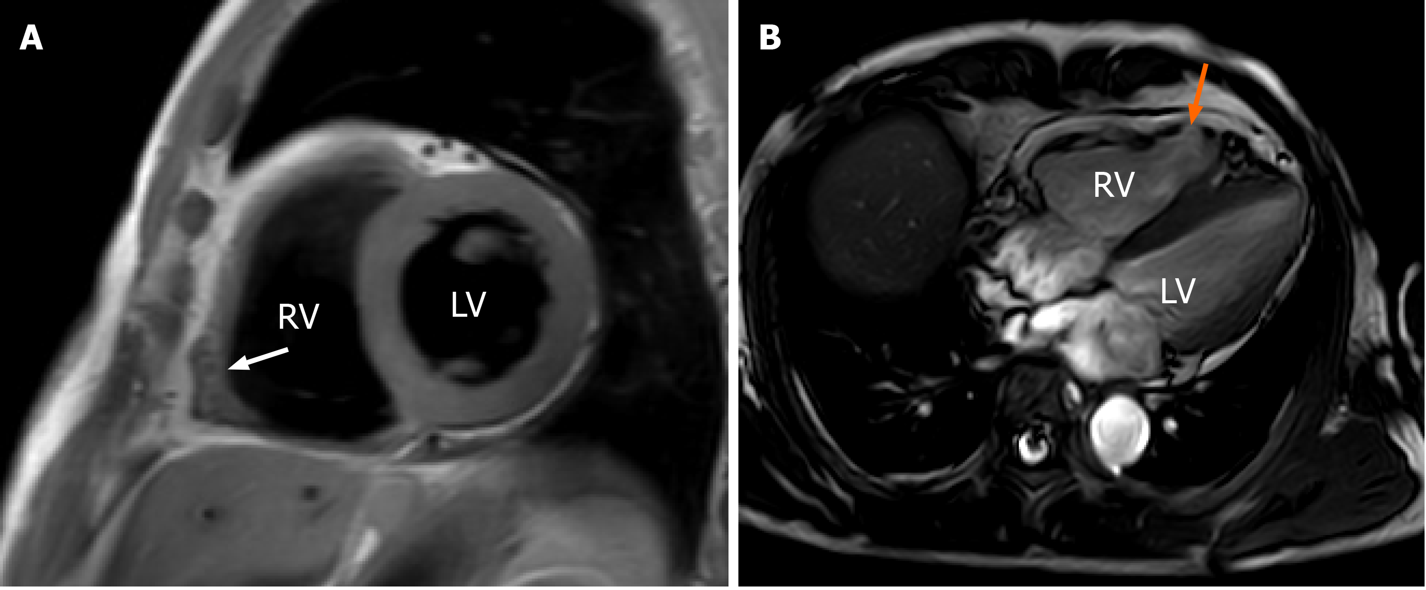
Figure 3 Cardiac magnetic resonance imaging in different views showed right ventricular free wall thinning, right ventricular dilatation, fibrofatty infiltration and regional right ventricular aneurysm.
A: Fibrofatty infiltration (white arrow); B: Regional right ventricular aneurysm (orange arrow). RV: Right ventricle; LV: Left ventricle.
- Citation: Wu HY, Cao YW, Gao TJ, Fu JL, Liang L. Arrhythmogenic right ventricular cardiomyopathy characterized by recurrent syncope during exercise: A case report. World J Clin Cases 2021; 9(16): 4095-4103
- URL: https://www.wjgnet.com/2307-8960/full/v9/i16/4095.htm
- DOI: https://dx.doi.org/10.12998/wjcc.v9.i16.4095


