Copyright
©The Author(s) 2021.
World J Clin Cases. Jun 6, 2021; 9(16): 4095-4103
Published online Jun 6, 2021. doi: 10.12998/wjcc.v9.i16.4095
Published online Jun 6, 2021. doi: 10.12998/wjcc.v9.i16.4095
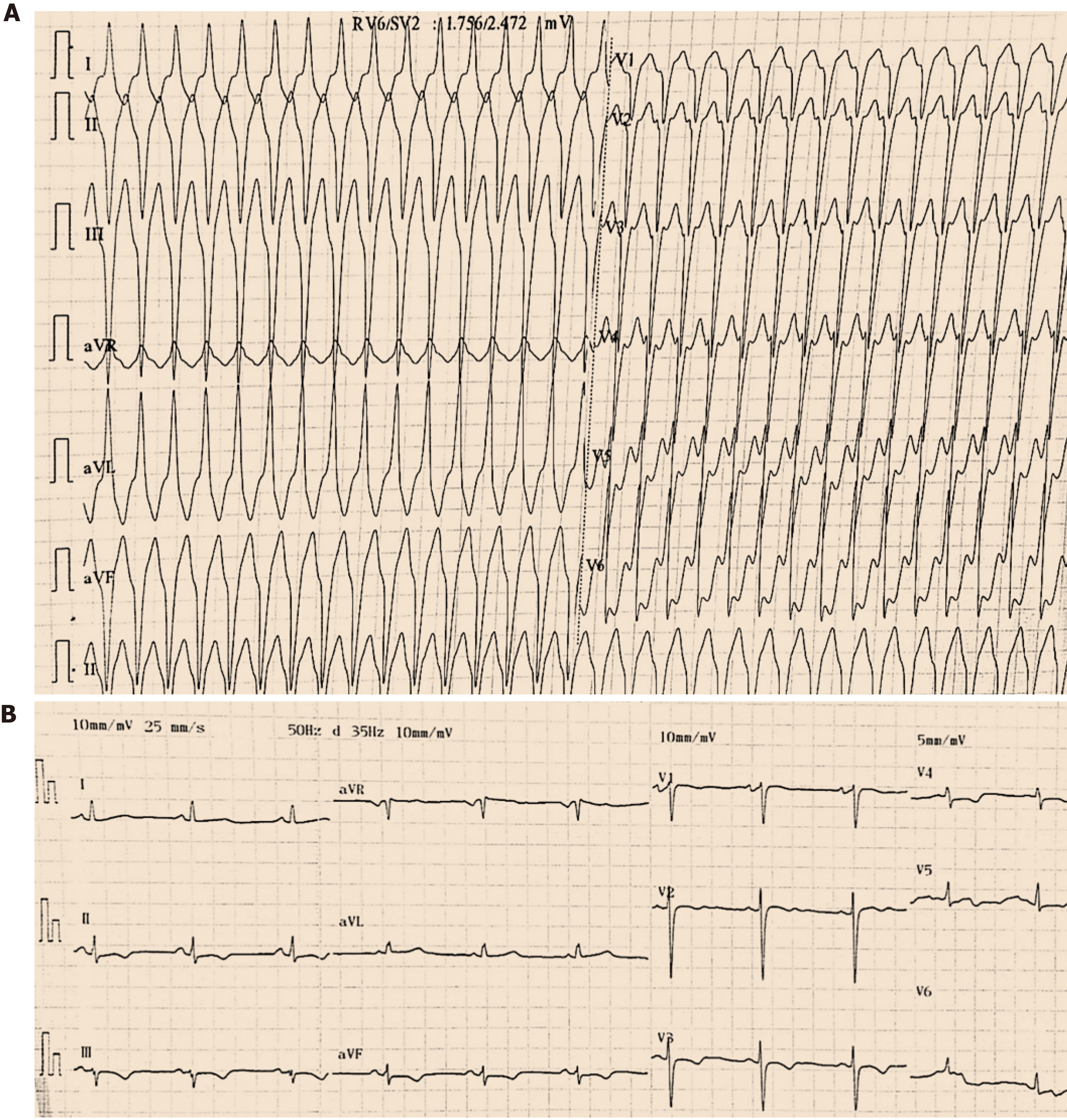
Figure 1 Twelve-lead electrocardiogram findings at admission and after drug cardioversion.
A: An electrocardiogram at admission revealed ventricular tachycardia (192 beats per minute) with a superior axis (positive QRS in lead aVL and negative QRS in leads II, III, and aVF), indicating an origin in the inferior wall of the right ventricle; B: An electrocardiogram after drug cardioversion showed a regular sinus rhythm at 65 beats per minute with negative T waves and a delayed S-wave upstroke (60 ms) from leads V1 to V4.
- Citation: Wu HY, Cao YW, Gao TJ, Fu JL, Liang L. Arrhythmogenic right ventricular cardiomyopathy characterized by recurrent syncope during exercise: A case report. World J Clin Cases 2021; 9(16): 4095-4103
- URL: https://www.wjgnet.com/2307-8960/full/v9/i16/4095.htm
- DOI: https://dx.doi.org/10.12998/wjcc.v9.i16.4095


