Copyright
©The Author(s) 2021.
World J Clin Cases. Jun 6, 2021; 9(16): 4072-4080
Published online Jun 6, 2021. doi: 10.12998/wjcc.v9.i16.4072
Published online Jun 6, 2021. doi: 10.12998/wjcc.v9.i16.4072
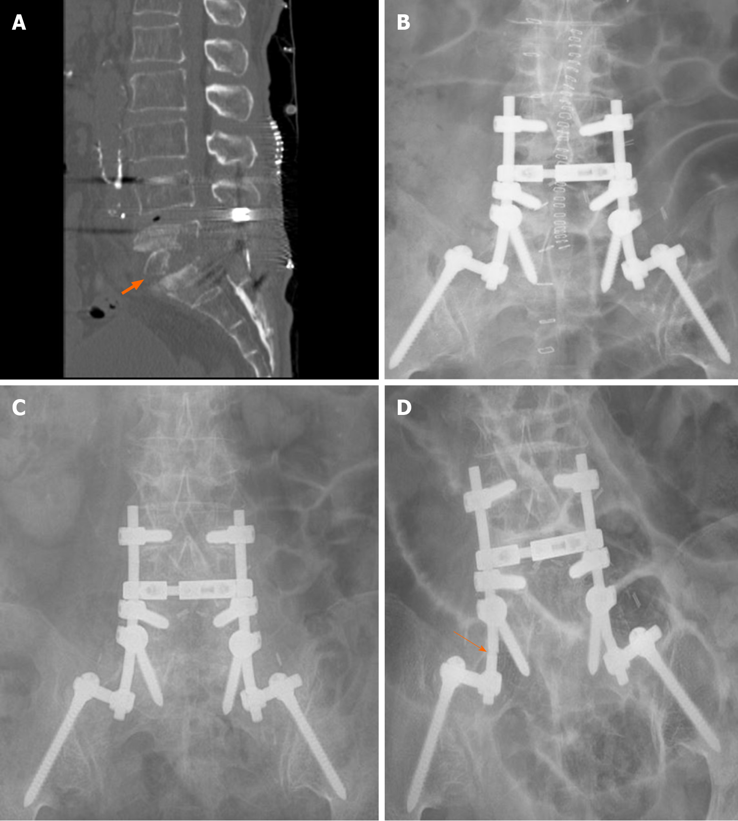
Figure 4 Follow-up spinal computed tomography and simple X-ray images after second surgery.
A: 1-wk postoperative spinal computed tomography shows bony erosion and autologous bone graft harvested from iliac crest (thick arrow); B: 1-wk postoperative simple X-rays; C: 3-year postoperative simple X-rays; D: 10-year postoperative simple X-rays. Ten-year postoperative simple X-ray revealed fracture of rod (thin arrow).
- Citation: Kim C, Lee S, Kim J. Spinal epidural abscess due to coinfection of bacteria and tuberculosis: A case report. World J Clin Cases 2021; 9(16): 4072-4080
- URL: https://www.wjgnet.com/2307-8960/full/v9/i16/4072.htm
- DOI: https://dx.doi.org/10.12998/wjcc.v9.i16.4072


