Copyright
©The Author(s) 2021.
World J Clin Cases. Jun 6, 2021; 9(16): 4016-4023
Published online Jun 6, 2021. doi: 10.12998/wjcc.v9.i16.4016
Published online Jun 6, 2021. doi: 10.12998/wjcc.v9.i16.4016
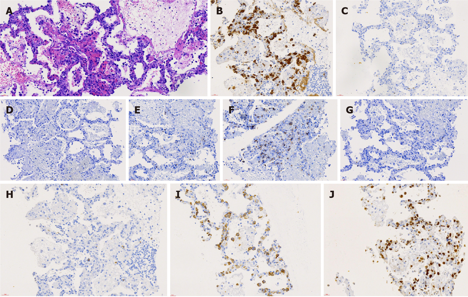
Figure 2 Pathological images of lung tissue biopsy at day 5 of hospitalization.
A: Hematoxylin-eosin staining showed the alveolar structure of the lung tissue with massive infiltration by large anaplastic lymphocytes in the alveolar septum with scattered neutrophils. Magnification, 400 ×; B-J: Immunostaining revealed that the tumor cells were ALK(+) (anaplastic lymphoma kinase) (B), CD3(-) (C), CD4(-) (D), CD5(-) (E), CD7(+) (F), CD8(-) (G), CD20(-) (H), CD30 (+) (I), and Ki-67(+) (J). Magnification, 400 ×.
- Citation: Jiang JH, Zhang CL, Wu QL, Liu YH, Wang XQ, Wang XL, Fang BM. Rapidly progressing primary pulmonary lymphoma masquerading as lung infectious disease: A case report and review of the literature. World J Clin Cases 2021; 9(16): 4016-4023
- URL: https://www.wjgnet.com/2307-8960/full/v9/i16/4016.htm
- DOI: https://dx.doi.org/10.12998/wjcc.v9.i16.4016


