Copyright
©The Author(s) 2021.
World J Clin Cases. Jun 6, 2021; 9(16): 3996-4000
Published online Jun 6, 2021. doi: 10.12998/wjcc.v9.i16.3996
Published online Jun 6, 2021. doi: 10.12998/wjcc.v9.i16.3996
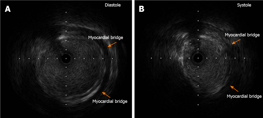
Figure 2 Images of intravascular ultrasound.
A: Intravascular ultrasound showed the coronary artery aneurysm and myocardial bridge during diastole; B: Intravascular ultrasound showed that the coronary artery aneurysm was compressed by the myocardial bridge during systole. The arrows indicate the location of the myocardial bridge.
- Citation: Ye Z, Dong XF, Yan YM, Luo YK. Coronary artery aneurysm combined with myocardial bridge: A case report. World J Clin Cases 2021; 9(16): 3996-4000
- URL: https://www.wjgnet.com/2307-8960/full/v9/i16/3996.htm
- DOI: https://dx.doi.org/10.12998/wjcc.v9.i16.3996


