Copyright
©The Author(s) 2021.
World J Clin Cases. Jun 6, 2021; 9(16): 3988-3995
Published online Jun 6, 2021. doi: 10.12998/wjcc.v9.i16.3988
Published online Jun 6, 2021. doi: 10.12998/wjcc.v9.i16.3988
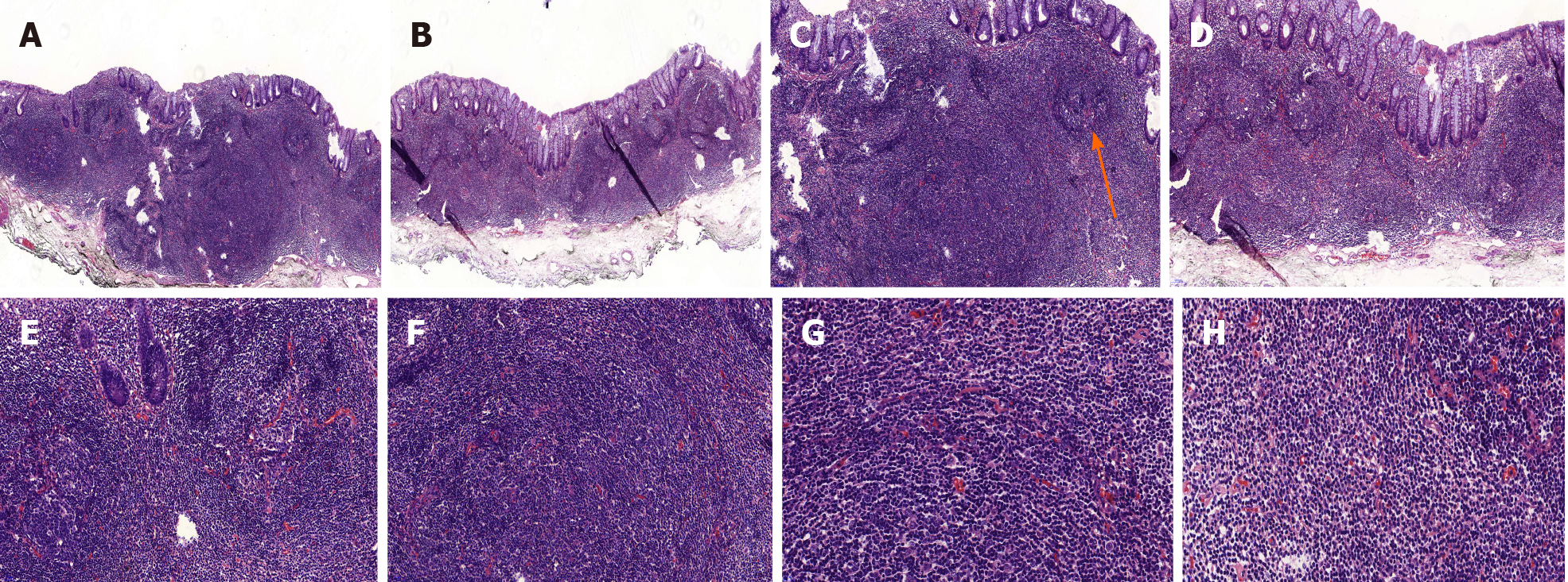
Figure 2 Histopathological findings (hematoxylin and eosin staining).
Diffusely hyperplastic lymphoid tissue could be observed in the lamina propria with visible lymphoid follicle structures (orange arrow, C). There were a large number of lymphoid cells around the lymphoid follicles that had clear cytoplasm and similar sizes. A and B: Magnification 50 ×; C and D: Magnification 100 ×; E and F: Magnification 200 ×; G and H Magnification 400 ×.
- Citation: Wei YL, Min CC, Ren LL, Xu S, Chen YQ, Zhang Q, Zhao WJ, Zhang CP, Yin XY. Laterally spreading tumor-like primary rectal mucosa-associated lymphoid tissue lymphoma: A case report. World J Clin Cases 2021; 9(16): 3988-3995
- URL: https://www.wjgnet.com/2307-8960/full/v9/i16/3988.htm
- DOI: https://dx.doi.org/10.12998/wjcc.v9.i16.3988


