Copyright
©The Author(s) 2021.
World J Clin Cases. Jun 6, 2021; 9(16): 3988-3995
Published online Jun 6, 2021. doi: 10.12998/wjcc.v9.i16.3988
Published online Jun 6, 2021. doi: 10.12998/wjcc.v9.i16.3988
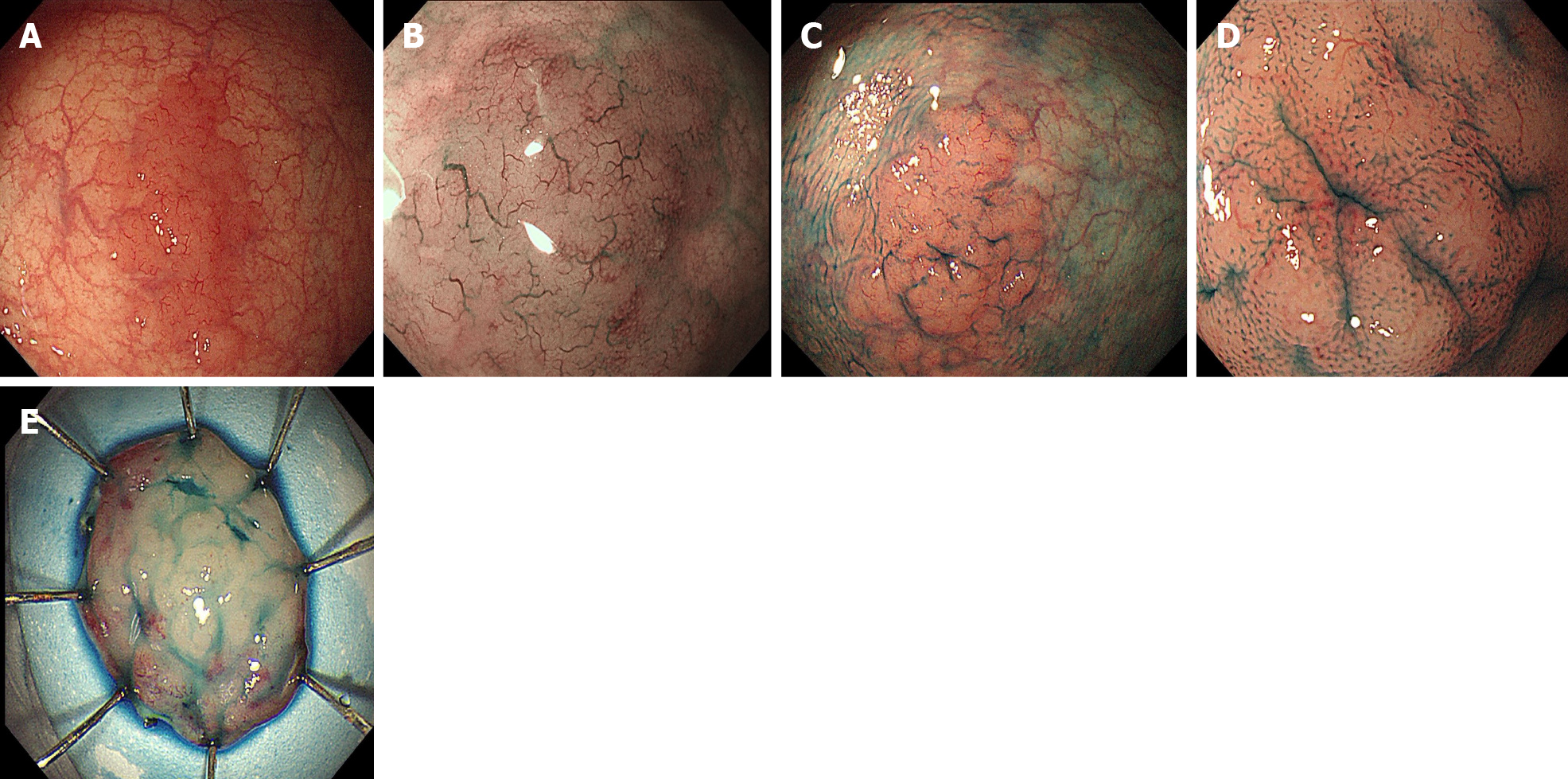
Figure 1 Endoscopic findings.
A: A laterally spreading tumor-like elevated lesion was observed on white light endoscopy in the rectum; B: Narrow band imaging showed enlarged branch-like vessels on the surface of the lesion; C and D: Indigo carmine staining made the lesion margin clearer (C), and pit pattern II was observed on magnifying endoscopy (D); E: On assessment of the endoscopic submucosal dissection specimen, a slightly elevated, laterally spreading tumor measuring 25 mm × 20 mm was identified in the mucosal layer.
- Citation: Wei YL, Min CC, Ren LL, Xu S, Chen YQ, Zhang Q, Zhao WJ, Zhang CP, Yin XY. Laterally spreading tumor-like primary rectal mucosa-associated lymphoid tissue lymphoma: A case report. World J Clin Cases 2021; 9(16): 3988-3995
- URL: https://www.wjgnet.com/2307-8960/full/v9/i16/3988.htm
- DOI: https://dx.doi.org/10.12998/wjcc.v9.i16.3988


