Copyright
©The Author(s) 2021.
World J Clin Cases. Jun 6, 2021; 9(16): 3979-3987
Published online Jun 6, 2021. doi: 10.12998/wjcc.v9.i16.3979
Published online Jun 6, 2021. doi: 10.12998/wjcc.v9.i16.3979
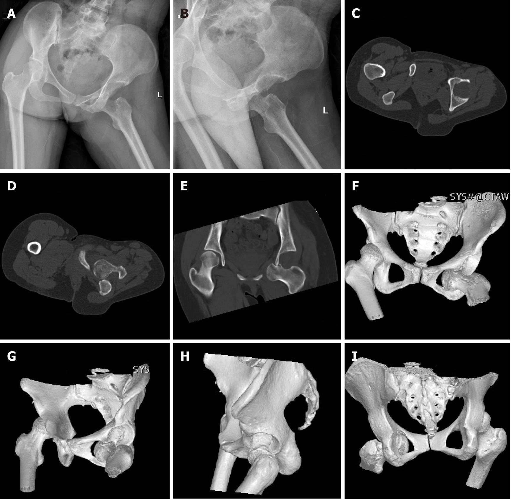
Figure 2 Preoperative plain radiographs and computed tomography scans.
A and B: Plain radiographs at the first examination showing obturator dislocation of the left hip; C-I: Axial, coronal, and three-dimensional reconstructed computed tomography images showing that the femoral head lay anteriorly and inferiorly to the obturator foramen, with impaction fracture at the superolateral aspect of the left femoral head without associated fracture of the acetabulum.
- Citation: Li WZ, Wang JJ, Ni JD, Song DY, Ding ML, Huang J, He GX. Old unreduced obturator dislocation of the hip: A case report. World J Clin Cases 2021; 9(16): 3979-3987
- URL: https://www.wjgnet.com/2307-8960/full/v9/i16/3979.htm
- DOI: https://dx.doi.org/10.12998/wjcc.v9.i16.3979


