Copyright
©The Author(s) 2021.
World J Clin Cases. Jun 6, 2021; 9(16): 3936-3942
Published online Jun 6, 2021. doi: 10.12998/wjcc.v9.i16.3936
Published online Jun 6, 2021. doi: 10.12998/wjcc.v9.i16.3936
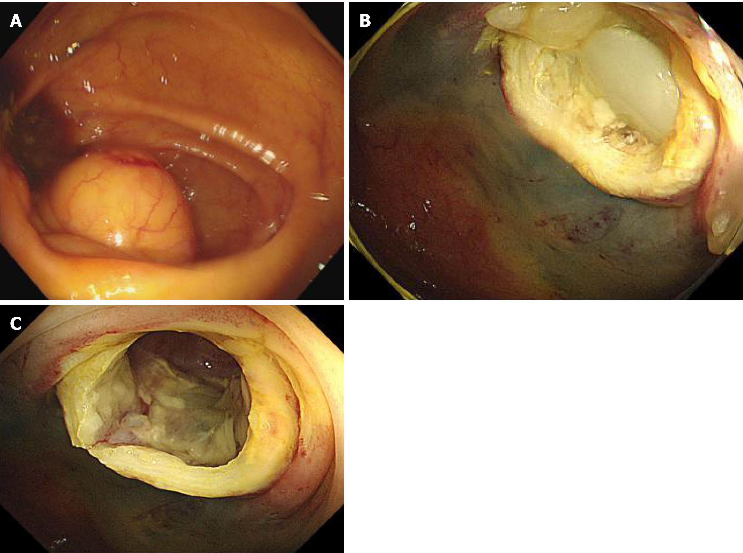
Figure 3 Colonoscopy.
A: A smooth-surfaced submucosal mass of the cecum with the appendiceal orifice in the center; B: A large amount of clear yellowish mucus flowing through the appendiceal orifice into the cecum after removing the submucosal mass; C: The inner wall of the appendix was smooth with no nodules.
- Citation: Wang TT, He JJ, Zhou PH, Chen WW, Chen CW, Liu J. Endoscopic diagnosis and treatment of an appendiceal mucocele: A case report. World J Clin Cases 2021; 9(16): 3936-3942
- URL: https://www.wjgnet.com/2307-8960/full/v9/i16/3936.htm
- DOI: https://dx.doi.org/10.12998/wjcc.v9.i16.3936


