Copyright
©The Author(s) 2021.
World J Clin Cases. Jun 6, 2021; 9(16): 3927-3935
Published online Jun 6, 2021. doi: 10.12998/wjcc.v9.i16.3927
Published online Jun 6, 2021. doi: 10.12998/wjcc.v9.i16.3927
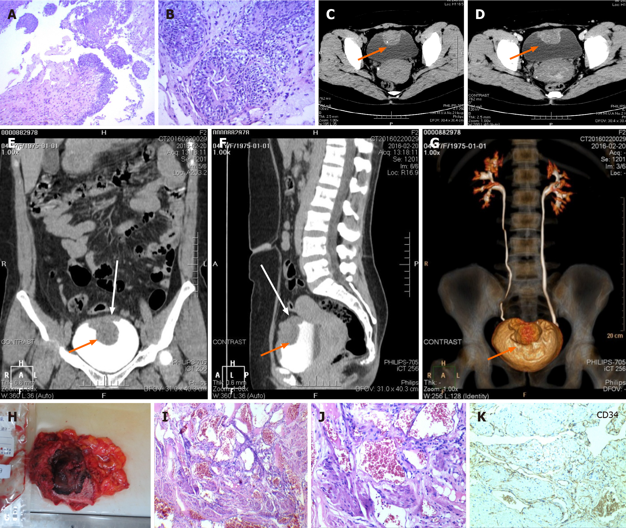
Figure 1 Cystoscopy and biopsy, multislice spiral computed tomography urography scanning, and histological and immunohistochemical characteristics of case 1.
A and B: The initial pathological report of cystoscopy and biopsy. Haematoxylin and eosin staining showed gland cystitis and local urothelial hyperplasia with nodule formation, but no clear cancer cells were found (A: 40 ×, B: 100 ×); C-G: Multislice spiral computed tomography urography of the urological system showed a 5.0 cm × 3.1 cm × 4.0 cm mass (orange arrow) arising from the superior and anterior wall of the urinary bladder with visible calcification and uneven enhancement; H: A specimen of the en bloc resected tumour. Macroscopically, it was a 10.0 cm × 7.0 cm × 4.0 cm partial cystectomy specimen, which on cut section showed a large, soft to firm haemorrhagic tumour mass measuring approximately 6.0 cm × 5.0 cm × 5.0 cm; and I-K: Histological and immunohistochemical characteristics of the en bloc resected tumour. Haematoxylin and eosin staining exhibited the urothelium of the bladder mucosa in the resected specimen and dilated thin-walled vessels in the detrusor muscle layer (I: 40 ×). The lesion was composed of small irregular angiomatous spaces lined by a simple layer of endothelial cells (J: 100 ×), and endothelial cells were CD34-positive (K: 100 ×).
- Citation: Zhao GC, Ke CX. Haemangiomas in the urinary bladder: Two case reports. World J Clin Cases 2021; 9(16): 3927-3935
- URL: https://www.wjgnet.com/2307-8960/full/v9/i16/3927.htm
- DOI: https://dx.doi.org/10.12998/wjcc.v9.i16.3927


