Copyright
©The Author(s) 2021.
World J Clin Cases. Jun 6, 2021; 9(16): 3895-3907
Published online Jun 6, 2021. doi: 10.12998/wjcc.v9.i16.3895
Published online Jun 6, 2021. doi: 10.12998/wjcc.v9.i16.3895
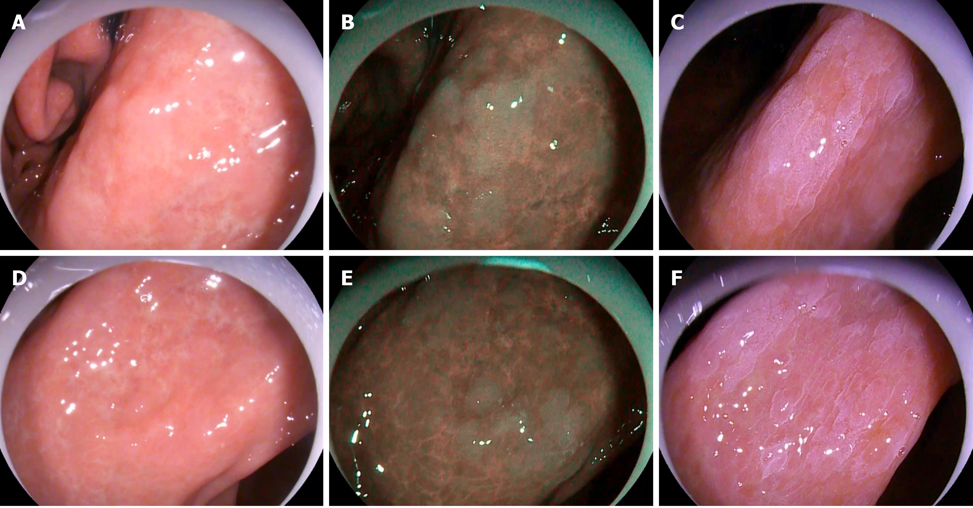
Figure 3 Appearance of gastric intestinal metaplasia under three different endoscopic methods.
A and D: Lesions as ash-colored nodular changes (white-light endoscopy); B and E: Bluish-whitish lesion area (optical-enhanced endoscopy, Mode 1); C and F: The clearer, whitish patches observed after spraying with acetic acid (acetic-acid chromoendoscopy).
- Citation: Song YH, Xu LD, Xing MX, Li KK, Xiao XG, Zhang Y, Li L, Xiao YJ, Qu YL, Wu HL. Comparison of white-light endoscopy, optical-enhanced and acetic-acid magnifying endoscopy for detecting gastric intestinal metaplasia: A randomized trial. World J Clin Cases 2021; 9(16): 3895-3907
- URL: https://www.wjgnet.com/2307-8960/full/v9/i16/3895.htm
- DOI: https://dx.doi.org/10.12998/wjcc.v9.i16.3895


