Copyright
©The Author(s) 2021.
World J Clin Cases. Jun 6, 2021; 9(16): 3895-3907
Published online Jun 6, 2021. doi: 10.12998/wjcc.v9.i16.3895
Published online Jun 6, 2021. doi: 10.12998/wjcc.v9.i16.3895
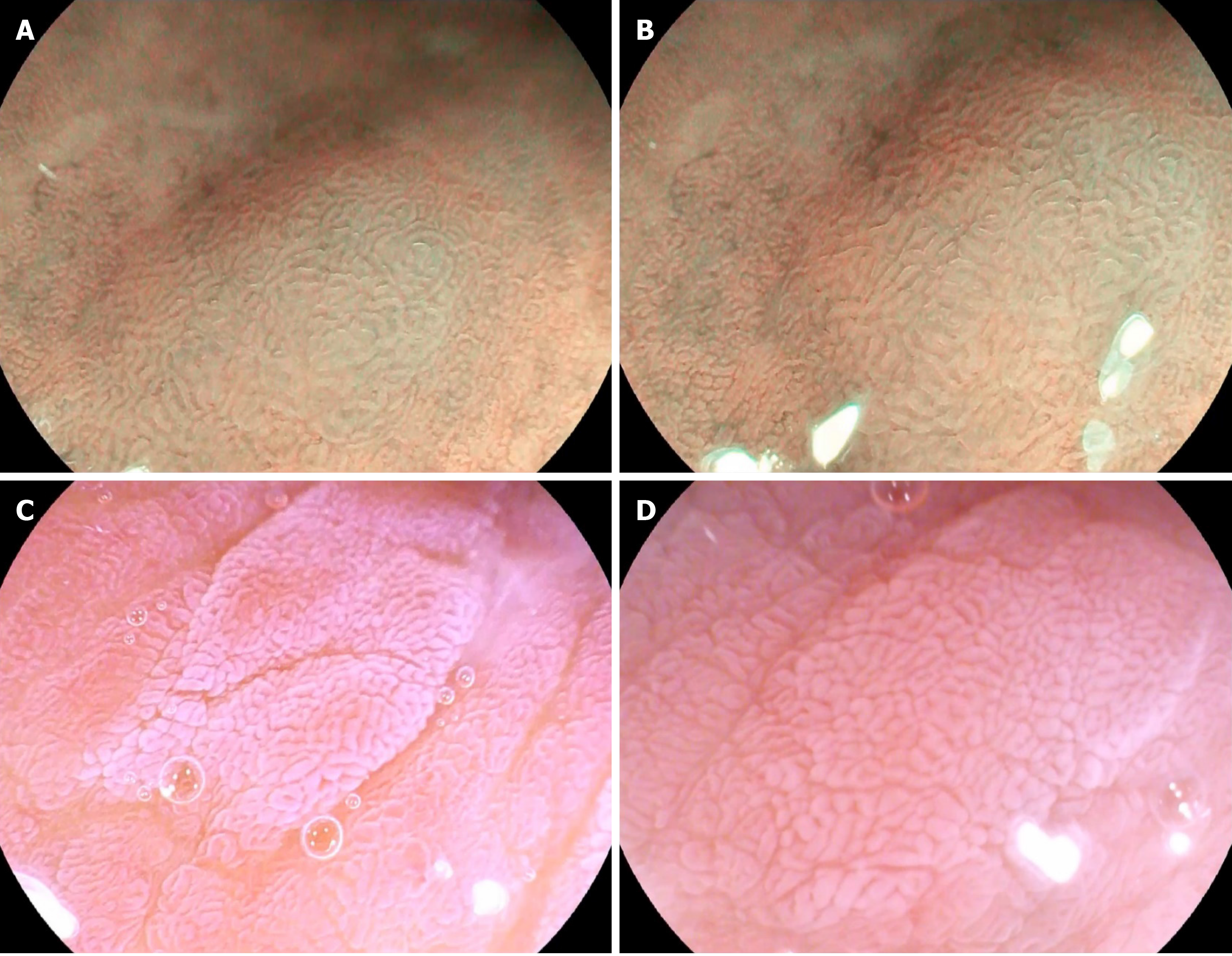
Figure 1 Endoscopic images in the magnifying mode.
A and B: In the optical-enhanced endoscopy Mode 1 and magnifying endoscopy image, light blue crest appears as blue-white lines visible on the epithelial surface; C and D: After spraying acetic acid, villous or cerebral gyrus-like structure, partial pits missing, and irregular arrangement are usually shown in magnifying mode.
- Citation: Song YH, Xu LD, Xing MX, Li KK, Xiao XG, Zhang Y, Li L, Xiao YJ, Qu YL, Wu HL. Comparison of white-light endoscopy, optical-enhanced and acetic-acid magnifying endoscopy for detecting gastric intestinal metaplasia: A randomized trial. World J Clin Cases 2021; 9(16): 3895-3907
- URL: https://www.wjgnet.com/2307-8960/full/v9/i16/3895.htm
- DOI: https://dx.doi.org/10.12998/wjcc.v9.i16.3895


