Copyright
©The Author(s) 2021.
World J Clin Cases. May 26, 2021; 9(15): 3779-3786
Published online May 26, 2021. doi: 10.12998/wjcc.v9.i15.3779
Published online May 26, 2021. doi: 10.12998/wjcc.v9.i15.3779
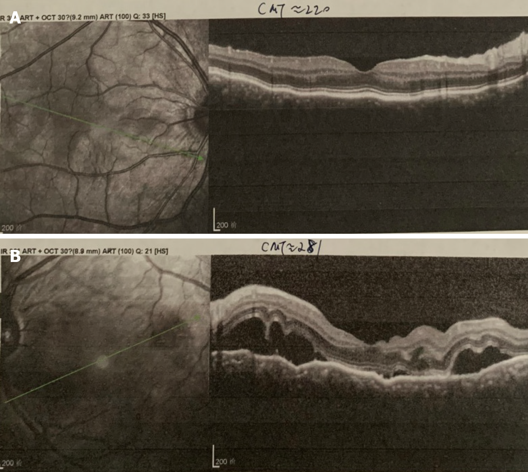
Figure 3 Optical coherence tomography examination before treatment.
A: Retinal pigment epithelial folds were found in the right eye; B: Optic disc edema and multiple serous retinal detachments were found in the left eye.
- Citation: Wen C, Duan H. Bilateral posterior scleritis presenting as acute primary angle closure: A case report. World J Clin Cases 2021; 9(15): 3779-3786
- URL: https://www.wjgnet.com/2307-8960/full/v9/i15/3779.htm
- DOI: https://dx.doi.org/10.12998/wjcc.v9.i15.3779


