Copyright
©The Author(s) 2021.
World J Clin Cases. May 26, 2021; 9(15): 3779-3786
Published online May 26, 2021. doi: 10.12998/wjcc.v9.i15.3779
Published online May 26, 2021. doi: 10.12998/wjcc.v9.i15.3779
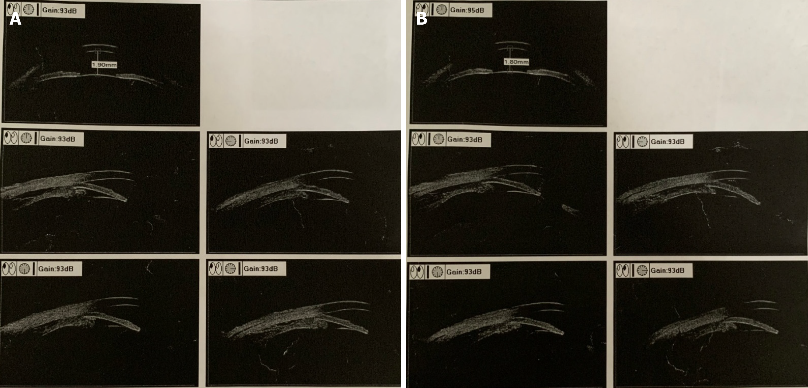
Figure 2 Ultrasound biomicroscopic examination before treatment.
The anterior chamber depths were 1.9 mm for the right eye and 1.8 mm for the left eye. Cyclodialysis, pronated ciliary process, and totally closed anterior chamber angle were present. A: Right eye; B: Left eye.
- Citation: Wen C, Duan H. Bilateral posterior scleritis presenting as acute primary angle closure: A case report. World J Clin Cases 2021; 9(15): 3779-3786
- URL: https://www.wjgnet.com/2307-8960/full/v9/i15/3779.htm
- DOI: https://dx.doi.org/10.12998/wjcc.v9.i15.3779


