Copyright
©The Author(s) 2021.
World J Clin Cases. May 26, 2021; 9(15): 3779-3786
Published online May 26, 2021. doi: 10.12998/wjcc.v9.i15.3779
Published online May 26, 2021. doi: 10.12998/wjcc.v9.i15.3779
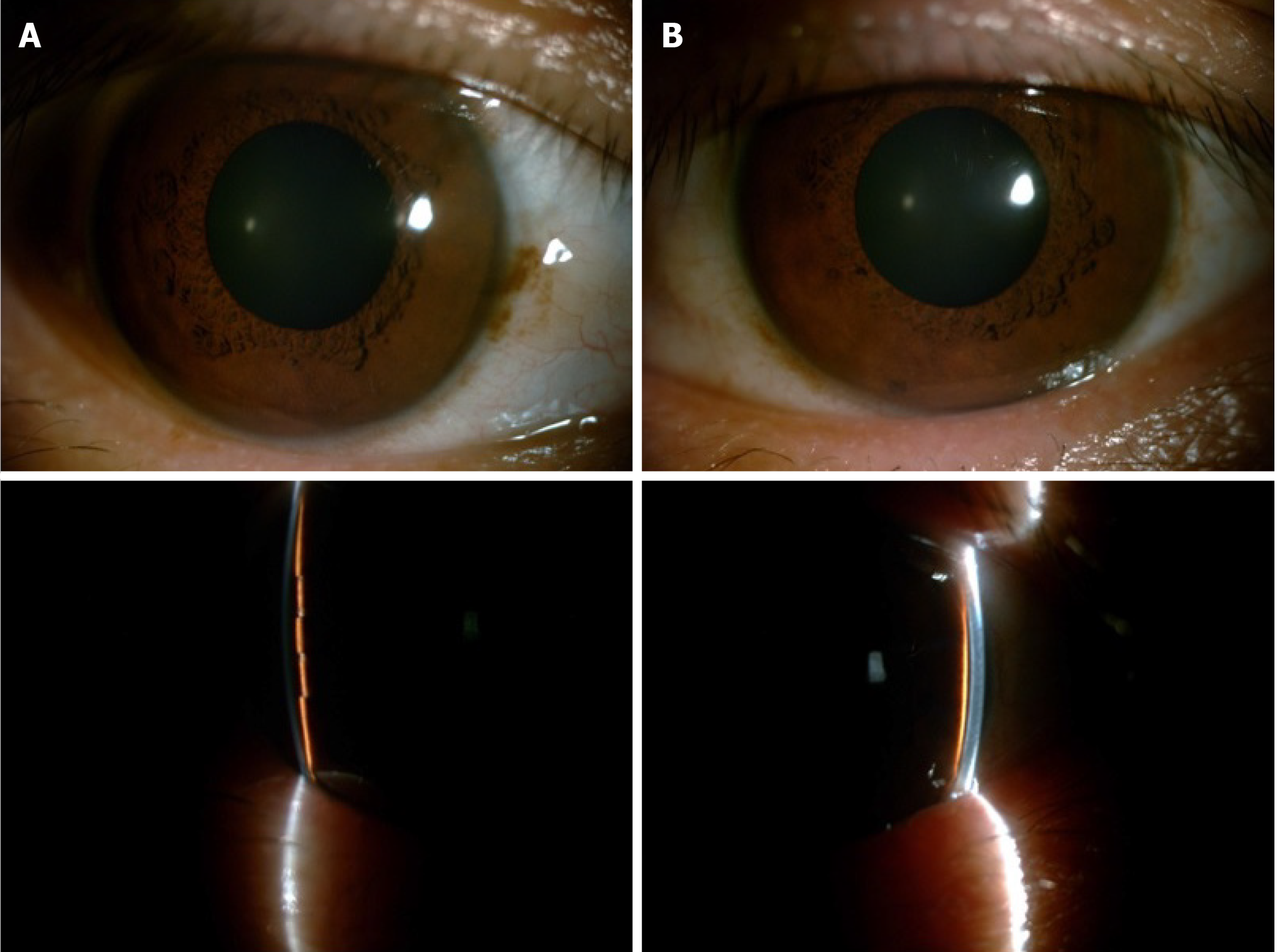
Figure 1 Anterior segment photography before treatment.
A quite shallow anterior chamber was present in both eyes and accompanied by mild cornea edema. The pupils were 5 mm, round, and symmetrical to each other. The lenses were transparent without dislocation. A: Right eye; B: Left eye.
- Citation: Wen C, Duan H. Bilateral posterior scleritis presenting as acute primary angle closure: A case report. World J Clin Cases 2021; 9(15): 3779-3786
- URL: https://www.wjgnet.com/2307-8960/full/v9/i15/3779.htm
- DOI: https://dx.doi.org/10.12998/wjcc.v9.i15.3779


