Copyright
©The Author(s) 2021.
World J Clin Cases. May 26, 2021; 9(15): 3773-3778
Published online May 26, 2021. doi: 10.12998/wjcc.v9.i15.3773
Published online May 26, 2021. doi: 10.12998/wjcc.v9.i15.3773
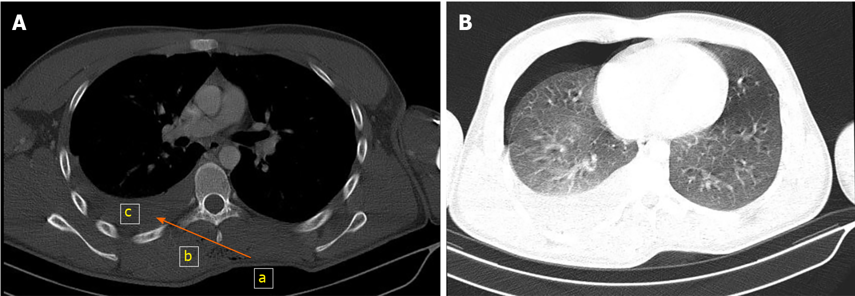
Figure 1 Pre-operative thoracic computed tomography.
A: Mediastinal window section of thoracic computed tomography: from a to b (level of the arrow). Penetration site of the sharp object (the pressure of dressing can be seen). A pneumoderma can be seen in the route taken by the sharp object from the left to right hemithorax; c: right hemithorax; B: Parenchymal window section of thoracic computed tomography: right hemopneumothorax.
- Citation: İşcan M. Contralateral hemopneumothorax after penetrating thoracic trauma: A case report. World J Clin Cases 2021; 9(15): 3773-3778
- URL: https://www.wjgnet.com/2307-8960/full/v9/i15/3773.htm
- DOI: https://dx.doi.org/10.12998/wjcc.v9.i15.3773


