Copyright
©The Author(s) 2021.
World J Clin Cases. May 26, 2021; 9(15): 3733-3740
Published online May 26, 2021. doi: 10.12998/wjcc.v9.i15.3733
Published online May 26, 2021. doi: 10.12998/wjcc.v9.i15.3733
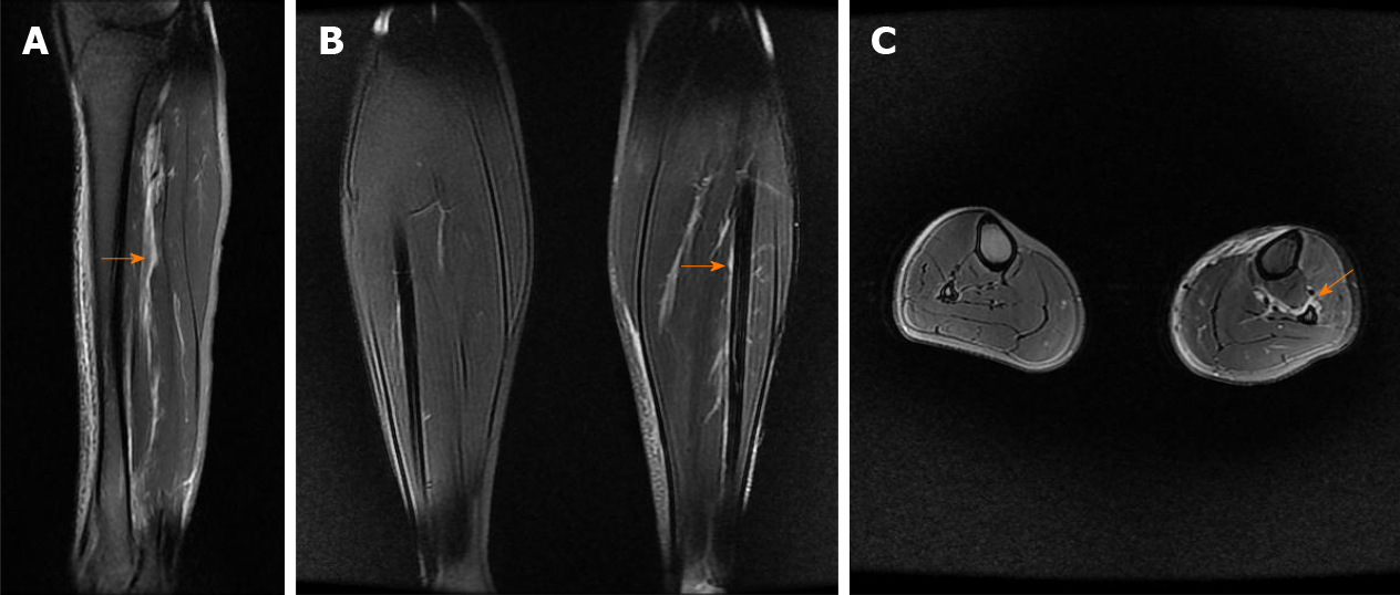
Figure 3 Calf magnetic resonance imaging.
A: Sagittal magnetic resonance imaging (MRI) showing high-signal intensity (orange arrow) indicating injury of the interosseous membrane (IOM); B: Coronal MRI showing IOM injury (orange arrow) compared with the contralateral uninjured calf; C: Axial MRI showing IOM rupture (orange arrow) compared with the contralateral uninjured calf, torn from the fibular interosseous crest.
- Citation: Liu GP, Li JG, Gong X, Li JM. Maisonneuve injury with no fibula fracture: A case report. World J Clin Cases 2021; 9(15): 3733-3740
- URL: https://www.wjgnet.com/2307-8960/full/v9/i15/3733.htm
- DOI: https://dx.doi.org/10.12998/wjcc.v9.i15.3733


