Copyright
©The Author(s) 2021.
World J Clin Cases. May 26, 2021; 9(15): 3726-3732
Published online May 26, 2021. doi: 10.12998/wjcc.v9.i15.3726
Published online May 26, 2021. doi: 10.12998/wjcc.v9.i15.3726
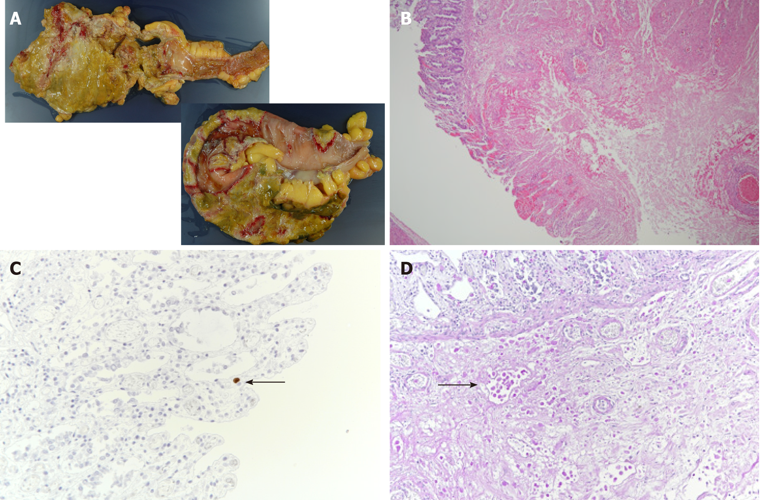
Figure 3 Macroscopic and histopathological findings during autopsy of a 68-year-old man who died after intestinal perforation.
A: Macroscopic findings revealing transmural intestinal necrosis and a “ragged appearance” in the rectum, descending colon, and sigmoid colon; B: Histopathological findings showing full-thickness necrosis of the intestinal wall from the mucosa to the serosa. Furthermore, severe infiltration of the necrotic tissue with ameba is visible (hematoxylin and eosin stain, × 4); C: A few intranuclear cytomegalovirus inclusions can be detected on immunohistochemical examination of the sigmoid colon (arrow, × 40); D: Entamoeba histolytica trophozoites are visible in the submucosa (periodic acid–Schiff stain, arrow, × 20).
- Citation: Shijubou N, Sumi T, Kamada K, Sawai T, Yamada Y, Ikeda T, Nakata H, Mori Y, Chiba H. Fulminant amebic colitis in a patient with concomitant cytomegalovirus infection after systemic steroid therapy: A case report . World J Clin Cases 2021; 9(15): 3726-3732
- URL: https://www.wjgnet.com/2307-8960/full/v9/i15/3726.htm
- DOI: https://dx.doi.org/10.12998/wjcc.v9.i15.3726


