Copyright
©The Author(s) 2021.
World J Clin Cases. May 26, 2021; 9(15): 3726-3732
Published online May 26, 2021. doi: 10.12998/wjcc.v9.i15.3726
Published online May 26, 2021. doi: 10.12998/wjcc.v9.i15.3726
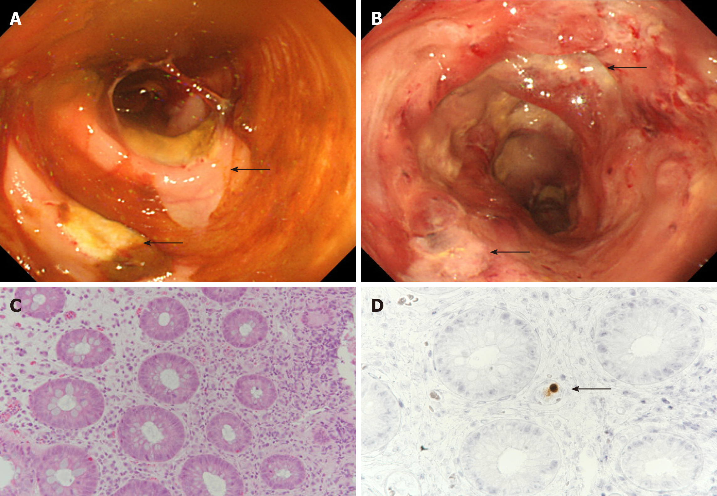
Figure 2 Colonoscopic and histopathological findings in a 68-year-old man who developed bloody diarrhea under glucocorticoid therapy.
A: Colonoscopy showing multiple large ulcers in the sigmoid colon (arrow) and active colitis throughout the colon; B: Colonoscopy also showing multiple large ulcers in the rectum (arrow); C: Neutrophilic and lymphocytic infiltration of the sigmoid colon’s mucosal interstitium (hematoxylin and eosin stain, × 20); D: Intranuclear inclusions of cytomegalovirus detected on immunohistochemical examination of the sigmoid colon’s interstitium (arrow, × 20).
- Citation: Shijubou N, Sumi T, Kamada K, Sawai T, Yamada Y, Ikeda T, Nakata H, Mori Y, Chiba H. Fulminant amebic colitis in a patient with concomitant cytomegalovirus infection after systemic steroid therapy: A case report . World J Clin Cases 2021; 9(15): 3726-3732
- URL: https://www.wjgnet.com/2307-8960/full/v9/i15/3726.htm
- DOI: https://dx.doi.org/10.12998/wjcc.v9.i15.3726


