Copyright
©The Author(s) 2021.
World J Clin Cases. May 26, 2021; 9(15): 3716-3725
Published online May 26, 2021. doi: 10.12998/wjcc.v9.i15.3716
Published online May 26, 2021. doi: 10.12998/wjcc.v9.i15.3716
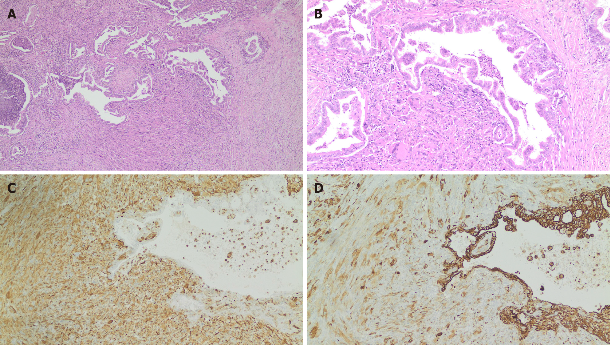
Figure 2 Histological examination and immunohistochemical staining.
A and B: Histological examination of the pancreatic neoplasm reveals infiltration by malignant cells displaying a glandular and spindle-cell pattern. Hematoxylin and eosin, 4 × (A); Hematoxylin and eosin, 10 × (B). Glandular component lined by atypical epithelium and sarcomatous spindle cell component with pleomorphic giant cells; C: Immunohistochemical staining for pan-cytokeratin, 10 × (C). Glandular and sarcomatoid components are positive for this epithelial marker; D: Immunohistochemical stain for vimentin, 10 × (D). Vimentin is the most common mesenchymal marker. The epithelial glandular component is negative, and the sarcomatoid component is strongly positive.
- Citation: Toledo PF, Berger Z, Carreño L, Cardenas G, Castillo J, Orellana O. Sarcomatoid carcinoma of the pancreas — a rare tumor with an uncommon presentation and course: A case report and review of literature. World J Clin Cases 2021; 9(15): 3716-3725
- URL: https://www.wjgnet.com/2307-8960/full/v9/i15/3716.htm
- DOI: https://dx.doi.org/10.12998/wjcc.v9.i15.3716


