Copyright
©The Author(s) 2021.
World J Clin Cases. May 26, 2021; 9(15): 3637-3643
Published online May 26, 2021. doi: 10.12998/wjcc.v9.i15.3637
Published online May 26, 2021. doi: 10.12998/wjcc.v9.i15.3637
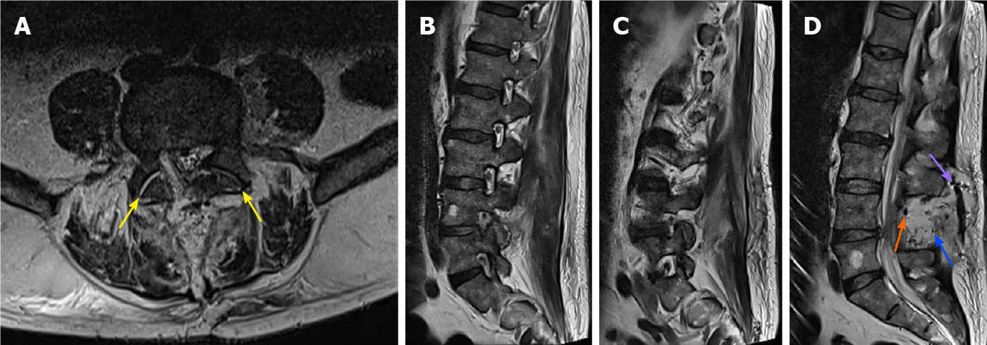
Figure 2 Magnetic resonance imaging scan on day 4 postoperatively.
A: T2-weighted axial magnetic resonance imaging (MRI) image showing a central decompression on L4-L5, with removal of the right-sided foraminal herniation. A fluid collection can be seen in both L4-L5 facet joints (yellow arrows); B and C: Right-sided and left-sided T2-weighted sagittal MRI images showing removal of the right-sided foraminal disc herniation; D: T2-weighted central sagittal MRI image showing a fluid collection (blue arrow), with small air collections (orange arrow) and a drain (purple arrow).
- Citation: Kerckhove MFV, Fiere V, Vieira TD, Bahroun S, Szadkowski M, d'Astorg H. Postoperative pain due to an occult spinal infection: A case report. World J Clin Cases 2021; 9(15): 3637-3643
- URL: https://www.wjgnet.com/2307-8960/full/v9/i15/3637.htm
- DOI: https://dx.doi.org/10.12998/wjcc.v9.i15.3637


