Copyright
©The Author(s) 2021.
World J Clin Cases. May 16, 2021; 9(14): 3356-3364
Published online May 16, 2021. doi: 10.12998/wjcc.v9.i14.3356
Published online May 16, 2021. doi: 10.12998/wjcc.v9.i14.3356
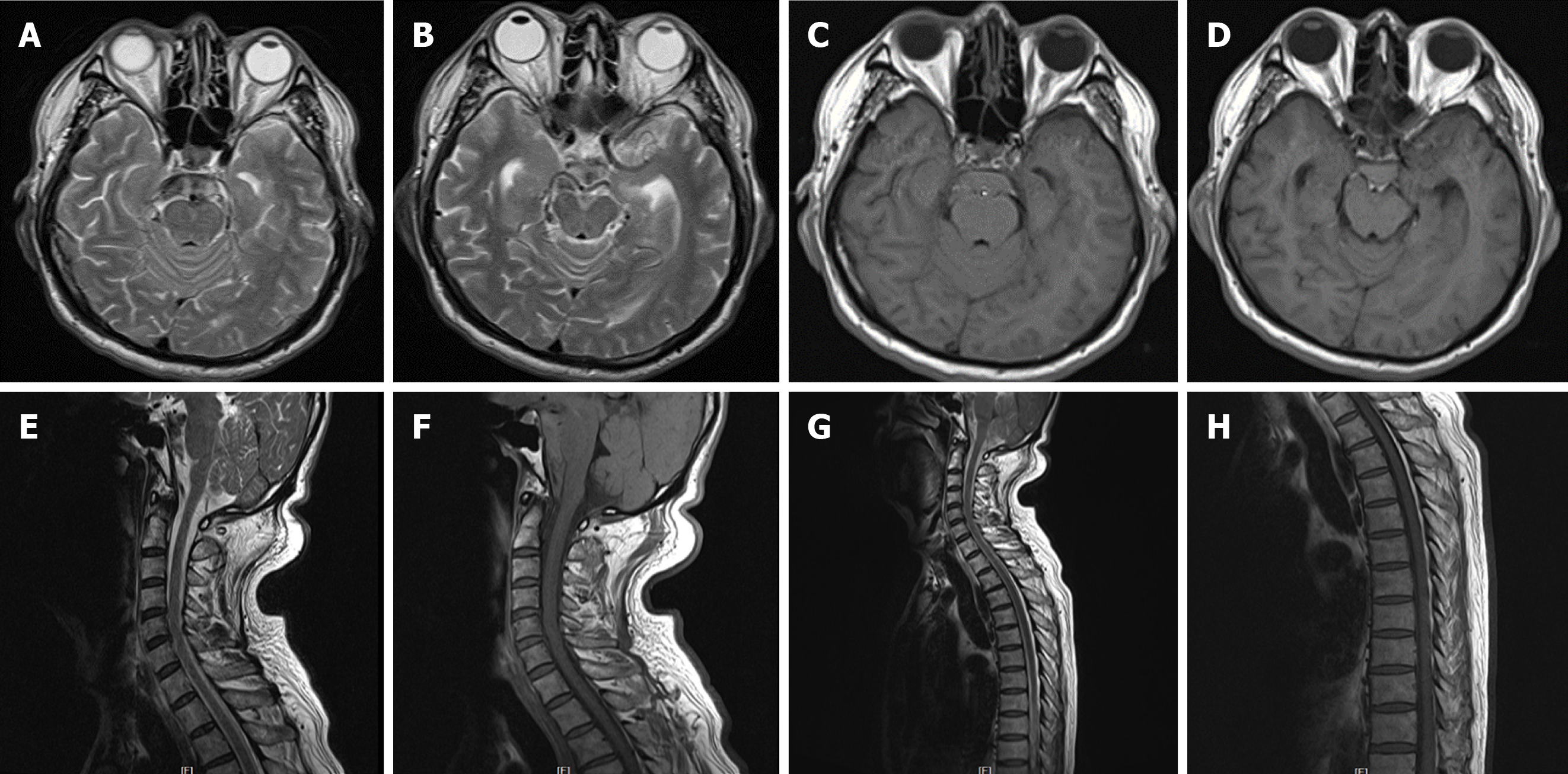
Figure 4 Magnetic resonance imaging of the brain, cervical spinal cord, and upper thoracic spinal cord performed the following day after second hemorrhage.
A and B: Axial T2 weighted imaging (T2WI) of the brain showed hyperintense signal; C and D: T1 weighted imaging (T1WI) exhibited equal intense signals in the perimesencephalic cisterns; E-H: Underlying structural abnormalities were not revealed. Sagittal T2WI and T1WI of the cervical and upper thoracic spinal cord were unremarkable.
- Citation: Li J, Fang X, Yu FC, Du B. Recurrent perimesencephalic nonaneurysmal subarachnoid hemorrhage within a short period of time: A case report. World J Clin Cases 2021; 9(14): 3356-3364
- URL: https://www.wjgnet.com/2307-8960/full/v9/i14/3356.htm
- DOI: https://dx.doi.org/10.12998/wjcc.v9.i14.3356


