Copyright
©The Author(s) 2021.
World J Clin Cases. May 16, 2021; 9(14): 3320-3326
Published online May 16, 2021. doi: 10.12998/wjcc.v9.i14.3320
Published online May 16, 2021. doi: 10.12998/wjcc.v9.i14.3320
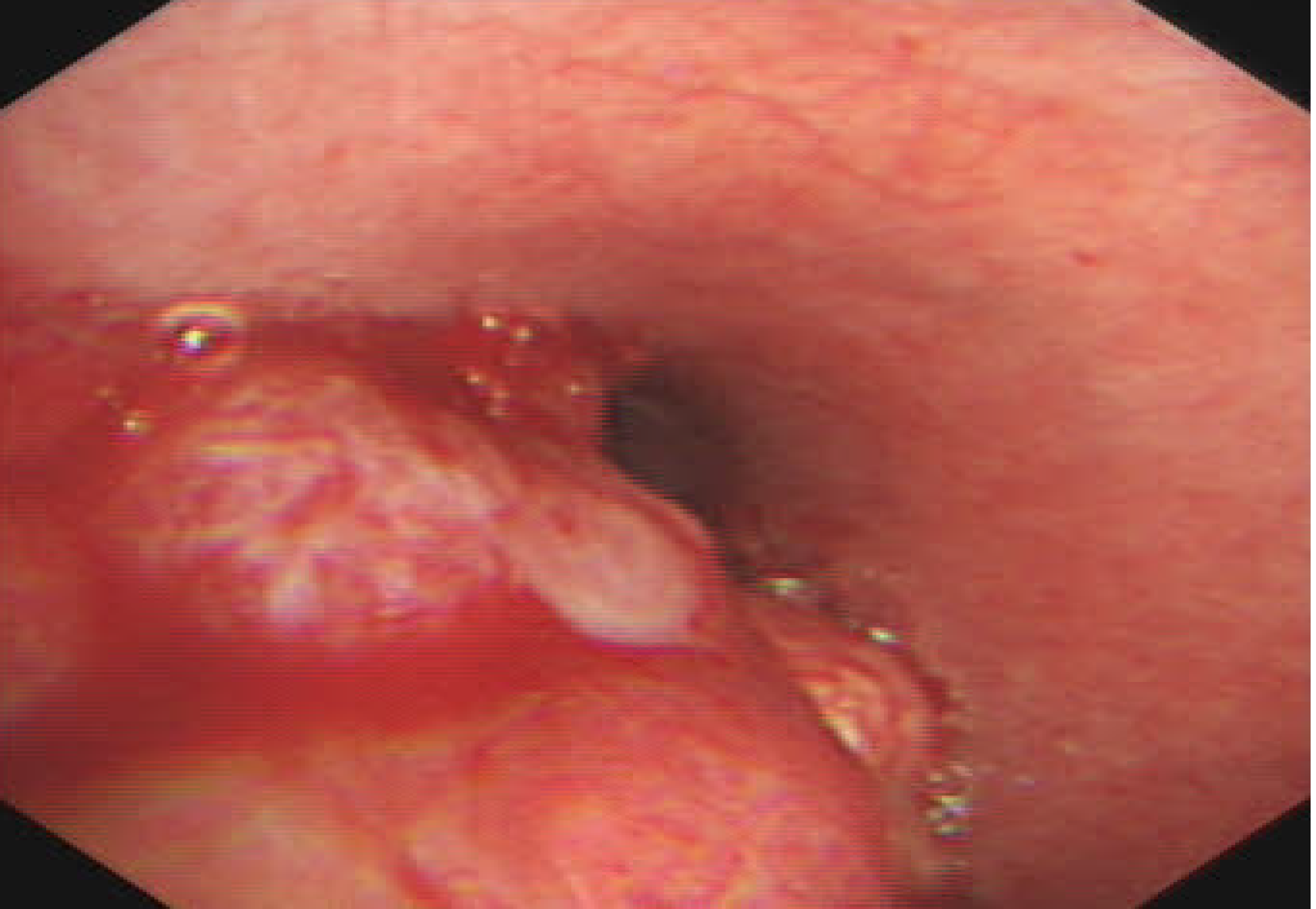
Figure 2 Bronchoscopic view of basal segment of the lower left lobe.
A yellow–white mass obstructed the entrance to the basal segment of the lower left lobe. Lateral to the entrance of the basal segment of the lower left lobe, neoplasms with multiple nodular ridges and superficial hyperemia were observed.
- Citation: Zhang Y, Zhang QP, Ji YQ, Xu J. Bronchial glomus tumor with calcification: A case report. World J Clin Cases 2021; 9(14): 3320-3326
- URL: https://www.wjgnet.com/2307-8960/full/v9/i14/3320.htm
- DOI: https://dx.doi.org/10.12998/wjcc.v9.i14.3320


