Copyright
©The Author(s) 2021.
World J Clin Cases. May 16, 2021; 9(14): 3320-3326
Published online May 16, 2021. doi: 10.12998/wjcc.v9.i14.3320
Published online May 16, 2021. doi: 10.12998/wjcc.v9.i14.3320
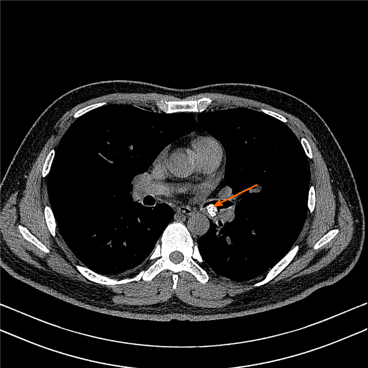
Figure 1 Chest computed tomography.
A 1.20 cm × 0.88 cm calcified nodular lesion on the compressed posterior wall of the lower left main bronchus (orange arrow).
- Citation: Zhang Y, Zhang QP, Ji YQ, Xu J. Bronchial glomus tumor with calcification: A case report. World J Clin Cases 2021; 9(14): 3320-3326
- URL: https://www.wjgnet.com/2307-8960/full/v9/i14/3320.htm
- DOI: https://dx.doi.org/10.12998/wjcc.v9.i14.3320


