Copyright
©The Author(s) 2021.
World J Clin Cases. May 6, 2021; 9(13): 3212-3218
Published online May 6, 2021. doi: 10.12998/wjcc.v9.i13.3212
Published online May 6, 2021. doi: 10.12998/wjcc.v9.i13.3212
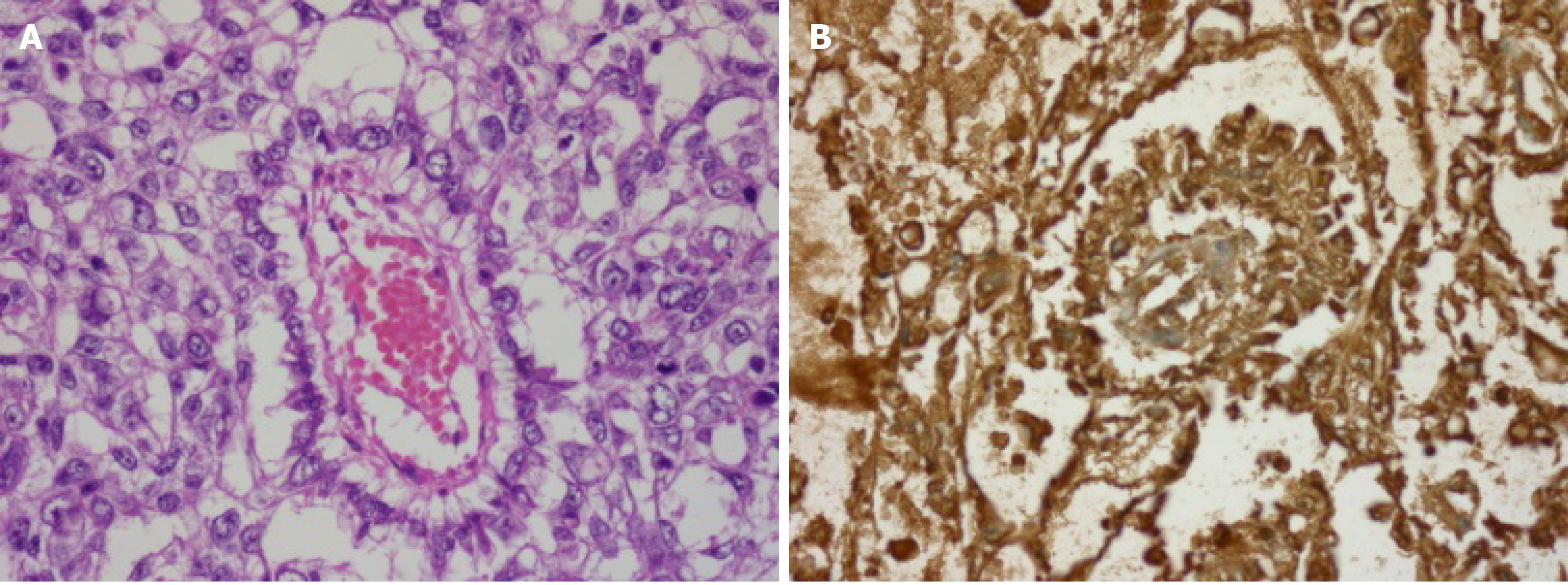
Figure 2 Pathology image.
A: Photomicrograph shows cuboidal-shaped tumor cells around central vessel forming the characteristic SchillerDuval body (hematoxylin and eosin, × 400); B: Immunohistochemical staining was positive for alpha-fetoprotein (original magnification × 400).
- Citation: Oh HK, Park SN, Kim BR. Laparoscopic uncontained power morcellation-induced dissemination of ovarian endodermal sinus tumors: A case report. World J Clin Cases 2021; 9(13): 3212-3218
- URL: https://www.wjgnet.com/2307-8960/full/v9/i13/3212.htm
- DOI: https://dx.doi.org/10.12998/wjcc.v9.i13.3212


