Copyright
©The Author(s) 2021.
World J Clin Cases. May 6, 2021; 9(13): 3185-3193
Published online May 6, 2021. doi: 10.12998/wjcc.v9.i13.3185
Published online May 6, 2021. doi: 10.12998/wjcc.v9.i13.3185
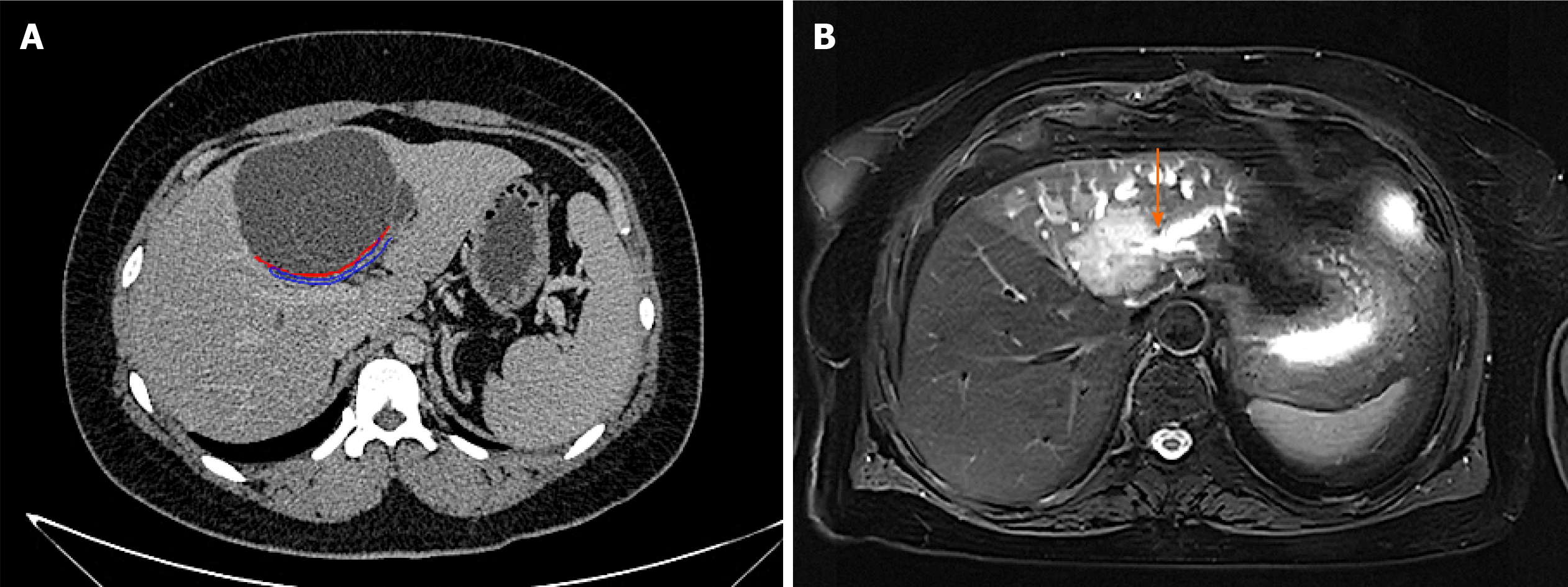
Figure 6 Differential diagnosis instructions.
A: Computed tomography image of mucinous cystic tumour of liver. It can be seen that the tumour does not communicate with bile duct. The orange arrow indicates the boundary of the tumour, and the blue arrow shows the bile duct compressed by the tumour; B: Magnetic resonance imaging image of cholangiocarcinoma. It can be seen that the boundary is not clear and smooth, and it invades into the liver, and the distal bile duct expands like a twig. The orange arrow indicates that the bile duct is out of shape and cut off at the lesion
- Citation: Yi D, Zhao LJ, Ding XB, Wang TW, Liu SY. Clinical characteristics of intrahepatic biliary papilloma: A case report. World J Clin Cases 2021; 9(13): 3185-3193
- URL: https://www.wjgnet.com/2307-8960/full/v9/i13/3185.htm
- DOI: https://dx.doi.org/10.12998/wjcc.v9.i13.3185


