Copyright
©The Author(s) 2021.
World J Clin Cases. May 6, 2021; 9(13): 3140-3146
Published online May 6, 2021. doi: 10.12998/wjcc.v9.i13.3140
Published online May 6, 2021. doi: 10.12998/wjcc.v9.i13.3140
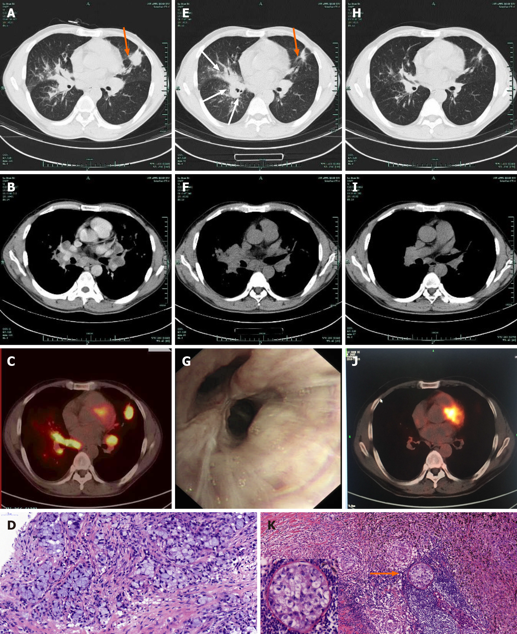
Figure 1 Thoracic computed tomography-scan, positron emission tomography- computed tomography, flexible bronchoscopy, and histopathological images.
A and B: Computed tomography (CT) image obtained on August 7, 2018, showing a 2.3 cm × 2.7 cm nodule (orange arrow) in the left superior lobe, small nodules of varying sizes in the left superior lobe and right lung, and enlarged bilateral hilar and mediastinal lymph nodes; C: Positron emission tomography-CT image obtained on August 8, 2018 showing hypermetabolic activity in the left superior lobe nodule and lymphadenopathy, suggestive of a neoplasm; D: Histopathological images obtained showing an adenocarcinoma sample from the superior lung nodule obtained by CT-guided pulmonary biopsy (hematoxylin and eosin, × 40); E and F: CT image obtained on September 7, 2018 after 30 d of treatment with crizotinib showed a dramatic decrease in the size of the left superior lobe nodule (orange arrow); however, the lesions in the right lung progressed (white arrows); G: Flexible bronchoscopy image showing multiple nodular infiltrations in the right middle lobe; H and I: CT image obtained on September 19, 2018 after 1 wk of methylprednisolone showing a significant response of all lesions in both lungs; J: Positron emission tomography-CT image obtained on November 15, 2018 showing a dramatic decrease in fluorodeoxyglucose uptake in pulmonary lesions; K: Histopathological image showing changes within the adenocarcinoma (orange arrow) with concomitant noncaseating granulomatous inflammation in samples from the parabronchial lymph nodes obtained by video-assisted thoracic surgery (hematoxylin and eosin, × 20).
- Citation: Chen X, Wang J, Han WL, Zhao K, Chen Z, Zhou JY, Shen YH. Sarcoidosis mimicking metastases in an echinoderm microtubule-associated protein-like 4 anaplastic lymphoma kinase positive non-small-lung cancer patient: A case report. World J Clin Cases 2021; 9(13): 3140-3146
- URL: https://www.wjgnet.com/2307-8960/full/v9/i13/3140.htm
- DOI: https://dx.doi.org/10.12998/wjcc.v9.i13.3140


