Copyright
©The Author(s) 2021.
World J Clin Cases. May 6, 2021; 9(13): 3102-3113
Published online May 6, 2021. doi: 10.12998/wjcc.v9.i13.3102
Published online May 6, 2021. doi: 10.12998/wjcc.v9.i13.3102
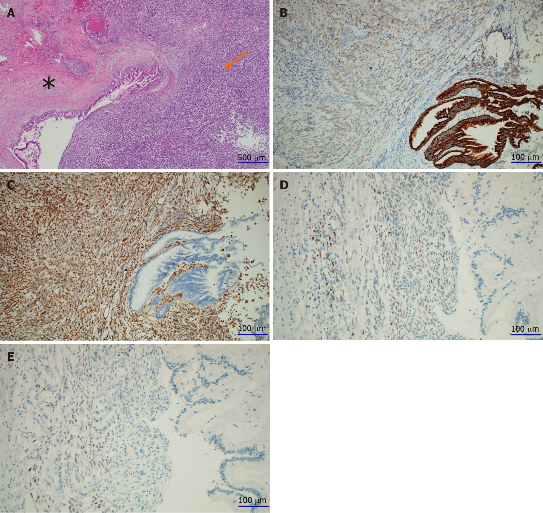
Figure 5 Histological examination of the lesion.
A: Microscopically, the tumor consisted of a central solid portion (arrow) and a thickened peripheral cystic wall (asterisk) on hematoxylin and eosin staining (magnification, × 40). Pathological findings showed that the tumors were adjacent to each other as two different components: sarcomatoid carcinoma (arrow) and mucinous cystic neoplasm (MCN) with high-grade dysplasia (asterisk); B and C: The MCN components were strongly and diffusely positive for pan-cytokeratin on immunostaining (magnification, × 200). The sarcomatoid carcinoma components were weakly positive for pan-cytokeratin (B) but strongly positive for vimentin on immunostaining (magnification, × 200) (C); D and E: The MCN had an ovarian-like stroma, which was immunohistochemically positive for the estrogen receptor (D), and the progesterone receptor (E) (magnification, × 200, respectively).
- Citation: Lim HJ, Kang HS, Lee JE, Min JH, Shin KS, You SK, Kim KH. Sarcomatoid carcinoma of the pancreas — multimodality imaging findings with serial imaging follow-up: A case report and review of literature. World J Clin Cases 2021; 9(13): 3102-3113
- URL: https://www.wjgnet.com/2307-8960/full/v9/i13/3102.htm
- DOI: https://dx.doi.org/10.12998/wjcc.v9.i13.3102


