Copyright
©The Author(s) 2021.
World J Clin Cases. May 6, 2021; 9(13): 3102-3113
Published online May 6, 2021. doi: 10.12998/wjcc.v9.i13.3102
Published online May 6, 2021. doi: 10.12998/wjcc.v9.i13.3102
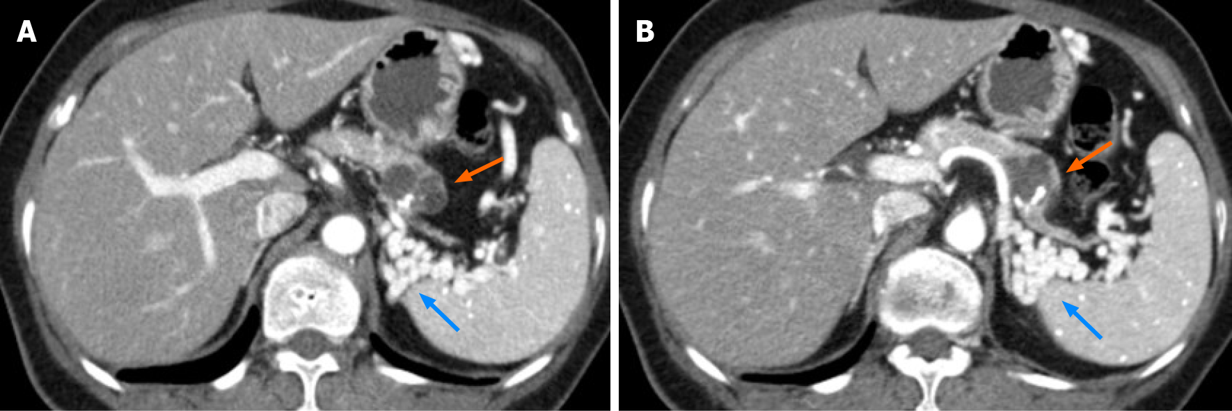
Figure 1 Initial computed tomography imaging of the abdomen.
A and B: Axial portal venous phase computed tomography images showing a 2.6 cm × 2.8 cm multilobulated cystic mass with an eccentric, relatively thick contrast-enhancing wall, and eccentric coarse calcification in the pancreatic body (orange arrows). No main pancreatic duct dilatation is observed. Upstream of the pancreatic parenchyma showed markedly atrophic changes, and the obliterated splenic vein was replaced with tortuous splenorenal collaterals (blue arrows).
- Citation: Lim HJ, Kang HS, Lee JE, Min JH, Shin KS, You SK, Kim KH. Sarcomatoid carcinoma of the pancreas — multimodality imaging findings with serial imaging follow-up: A case report and review of literature. World J Clin Cases 2021; 9(13): 3102-3113
- URL: https://www.wjgnet.com/2307-8960/full/v9/i13/3102.htm
- DOI: https://dx.doi.org/10.12998/wjcc.v9.i13.3102


