Copyright
©The Author(s) 2021.
World J Clin Cases. May 6, 2021; 9(13): 3090-3094
Published online May 6, 2021. doi: 10.12998/wjcc.v9.i13.3090
Published online May 6, 2021. doi: 10.12998/wjcc.v9.i13.3090
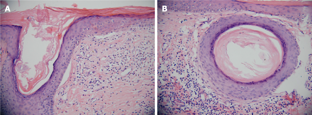
Figure 2 Histopathological findings from the left calf erosion (hematoxylin-eosin, original magnification × 20).
A: Hyperkeratosis and subepidermal cleft with moderate inflammatory infiltrate in the upper dermis showing scar tissue formation; B: The dermis revealed round keratin cysts with chronic inflammatory infiltrates in the upper dermis.
- Citation: Wang Z, Lin Y, Duan XW, Hang HY, Zhang X, Li LL. Misdiagnosed dystrophic epidermolysis bullosa pruriginosa: A case report. World J Clin Cases 2021; 9(13): 3090-3094
- URL: https://www.wjgnet.com/2307-8960/full/v9/i13/3090.htm
- DOI: https://dx.doi.org/10.12998/wjcc.v9.i13.3090


