Copyright
©The Author(s) 2021.
World J Clin Cases. May 6, 2021; 9(13): 3079-3089
Published online May 6, 2021. doi: 10.12998/wjcc.v9.i13.3079
Published online May 6, 2021. doi: 10.12998/wjcc.v9.i13.3079
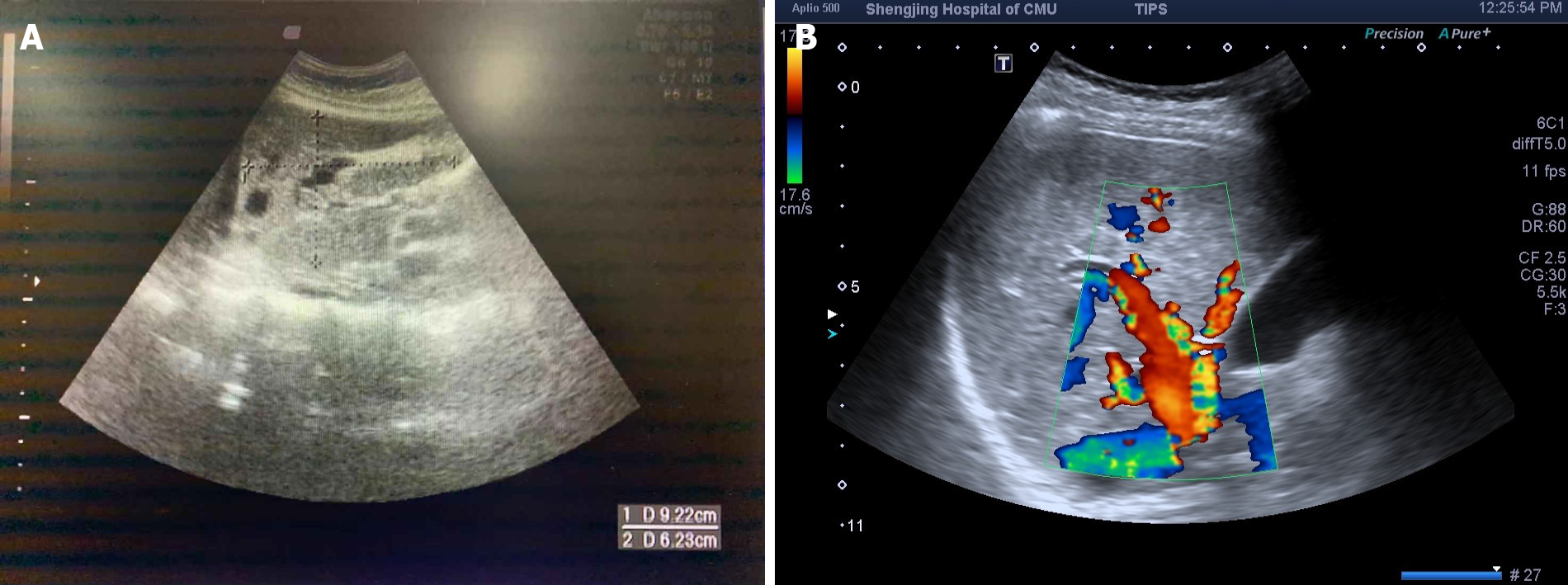
Figure 1 Liver ultrasound images of the proband and her relative.
A: The proband (β) showed vasodilation in the left lobe of the liver; B: The proband's relative (ε) showed hepatic artery dilatation, with a tortuous shape.
-
Citation: Wu J, Yuan Y, Wang X, Shao DY, Liu LG, He J, Li P. Pulmonary arterial hyper
tension in a patient with hereditary hemorrhagic telangiectasia and family gene analysis: A case report. World J Clin Cases 2021; 9(13): 3079-3089 - URL: https://www.wjgnet.com/2307-8960/full/v9/i13/3079.htm
- DOI: https://dx.doi.org/10.12998/wjcc.v9.i13.3079


