Copyright
©The Author(s) 2021.
World J Clin Cases. May 6, 2021; 9(13): 3063-3069
Published online May 6, 2021. doi: 10.12998/wjcc.v9.i13.3063
Published online May 6, 2021. doi: 10.12998/wjcc.v9.i13.3063
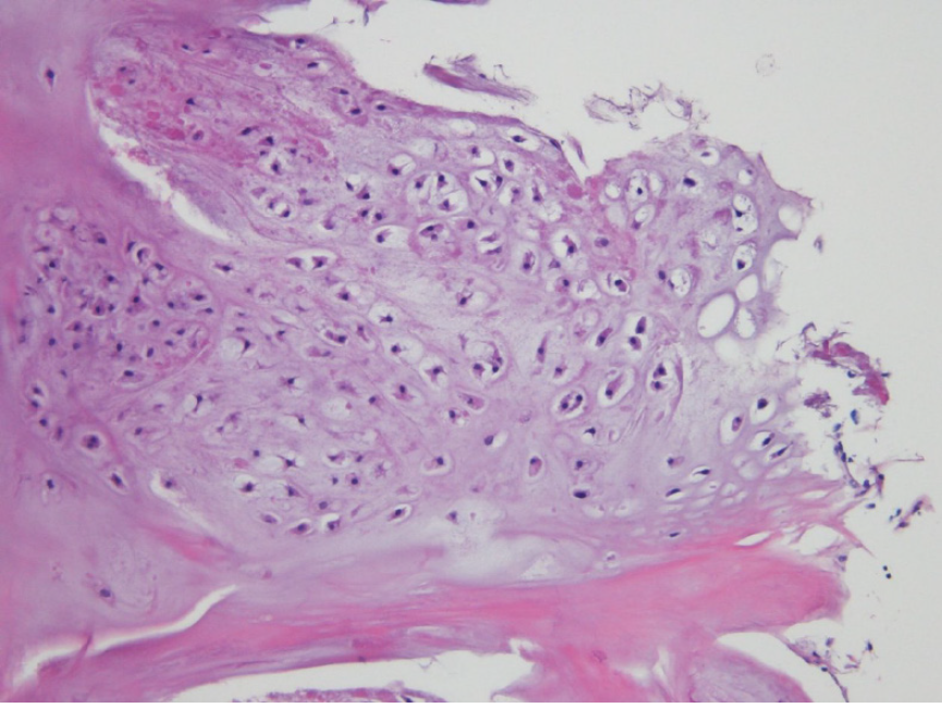
Figure 4 Histopathological image of the tumor.
Hypocellular, cytologically banal hyaline cartilage suggested chondroma and was located in the cortical bone. The sample was stained with hematoxylin-eosin and imaged at × 100 magnification.
- Citation: Yoshida Y, Anazawa U, Watanabe I, Hotta H, Aoyama R, Suzuki S, Nagura T. Intracortical chondroma of the metacarpal bone: A case report. World J Clin Cases 2021; 9(13): 3063-3069
- URL: https://www.wjgnet.com/2307-8960/full/v9/i13/3063.htm
- DOI: https://dx.doi.org/10.12998/wjcc.v9.i13.3063


