Copyright
©The Author(s) 2021.
World J Clin Cases. May 6, 2021; 9(13): 3063-3069
Published online May 6, 2021. doi: 10.12998/wjcc.v9.i13.3063
Published online May 6, 2021. doi: 10.12998/wjcc.v9.i13.3063
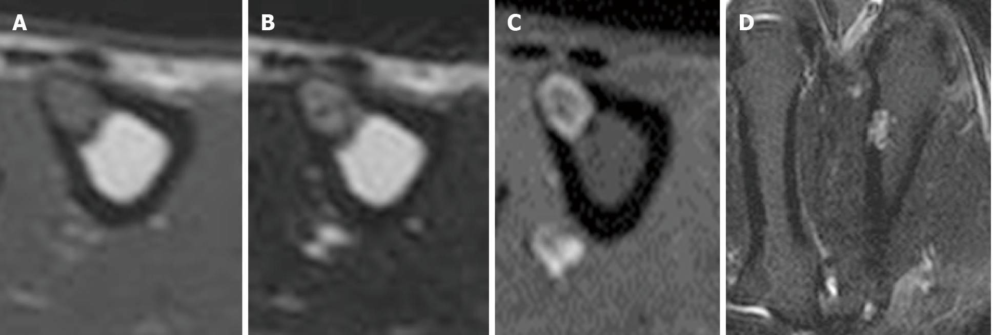
Figure 3 Magnetic resonance imaging of the second metacarpal bone.
A: Low signal intensity on axial T1-weighted images; B: High signal intensity on axial T2-weighted images; C: Axial contrast-enhanced magnetic resonance imaging. Only the tumor margin was enhanced; D: Coronal short tau inversion recovery magnetic resonance imaging.
- Citation: Yoshida Y, Anazawa U, Watanabe I, Hotta H, Aoyama R, Suzuki S, Nagura T. Intracortical chondroma of the metacarpal bone: A case report. World J Clin Cases 2021; 9(13): 3063-3069
- URL: https://www.wjgnet.com/2307-8960/full/v9/i13/3063.htm
- DOI: https://dx.doi.org/10.12998/wjcc.v9.i13.3063


