Copyright
©The Author(s) 2021.
World J Clin Cases. May 6, 2021; 9(13): 3048-3055
Published online May 6, 2021. doi: 10.12998/wjcc.v9.i13.3048
Published online May 6, 2021. doi: 10.12998/wjcc.v9.i13.3048
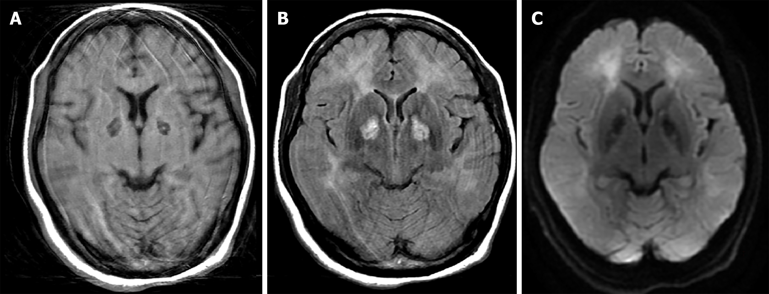
Figure 1 Brain magnetic resonance images of the patient.
A: Axial view of T1-weighted image shows low signal of bilateral globus pallidi, with a tiny high signal indicating minor hemorrhage; B: T2-Fluid-Attenuated Inversion Recovery image shows hyperintensities of bilateral globus pallidi and diffuse leukoencephalopathy in subcortical white matter of the bilateral frontal and occipital lobes; and C: Diffusion-weighted image shows increased diffusion signals. Artifacts were caused by the patient’s movement during image acquisition.
- Citation: Liu CC, Hsu CS, He HC, Cheng YY, Chang ST. Effects of intravascular laser phototherapy on delayed neurological sequelae after carbon monoxide intoxication as evaluated by brain perfusion imaging: A case report and review of the literature. World J Clin Cases 2021; 9(13): 3048-3055
- URL: https://www.wjgnet.com/2307-8960/full/v9/i13/3048.htm
- DOI: https://dx.doi.org/10.12998/wjcc.v9.i13.3048


