Copyright
©The Author(s) 2021.
World J Clin Cases. Apr 26, 2021; 9(12): 2937-2943
Published online Apr 26, 2021. doi: 10.12998/wjcc.v9.i12.2937
Published online Apr 26, 2021. doi: 10.12998/wjcc.v9.i12.2937
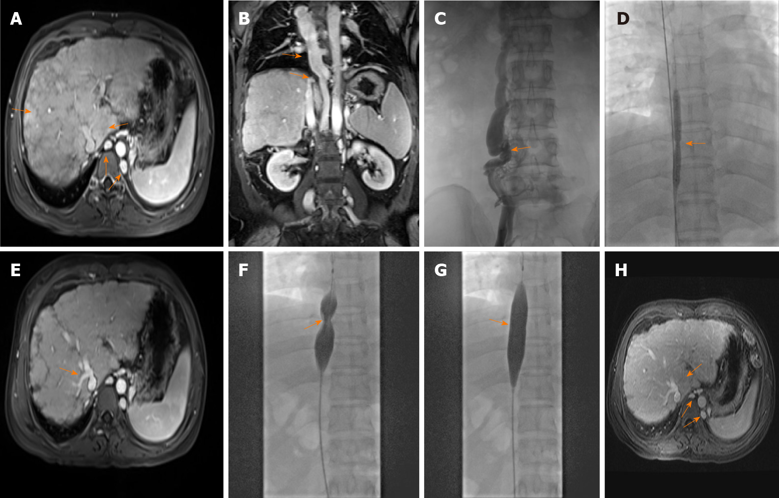
Figure 1 Imaging findings of Budd-Chiari syndrome with membranous obstruction of the inferior vena cava.
A: Magnetic resonance imaging (MRI) showed caudate lobe hypertrophy, cirrhosis, and dilated lumbar vein and hemiazygos veins on June 13, 2018; B: MRI revealed dilated azygos veins and narrowed inferior vena cava (IVC) on June 13, 2018; C: Venography of hepatic veins and IVC revealed complete occlusion of the IVC with the formation of numerous collateral branches on June 19, 2018; D: Balloon angioplasty was performed to recanalize the obstructed IVC on June 19, 2018; E: MRI revealed hepatic vein stenosis with ectopic tissue 3 mo after balloon angioplasty; F and G: Before and after balloon angioplasty of the IVC; H: Six months after balloon angioplasty, MRI revealed that dilation of the lumbar and hemiazygos veins and caudate lobe hypertrophy were improved.
- Citation: Ye QB, Huang QF, Luo YC, Wen YL, Chen ZK, Wei AL. Budd-Chiari syndrome associated with liver cirrhosis: A case report. World J Clin Cases 2021; 9(12): 2937-2943
- URL: https://www.wjgnet.com/2307-8960/full/v9/i12/2937.htm
- DOI: https://dx.doi.org/10.12998/wjcc.v9.i12.2937


