Copyright
©The Author(s) 2021.
World J Clin Cases. Apr 26, 2021; 9(12): 2908-2915
Published online Apr 26, 2021. doi: 10.12998/wjcc.v9.i12.2908
Published online Apr 26, 2021. doi: 10.12998/wjcc.v9.i12.2908
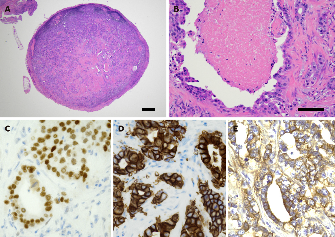
Figure 4 Histological analyses.
A and B: Biopsy of the lesion composed of atypical epithelioid cells within fibrous tissue. A nuclear pleomorphism and occasional mitoses are also noted. Scale bars represent 1000 μm (A) and 100 μm (B), respectively; C-E: Immunohistochemical staining for androgen receptor (C), human epidermal growth factor receptor 2 (D), and epithelial growth factor receptor (E).
- Citation: Uchihashi T, Kodama S, Sugauchi A, Hiraoka S, Hirose K, Usami Y, Tanaka S, Kogo M. Salivary duct carcinoma of the submandibular gland presenting a diagnostic challenge: A case report. World J Clin Cases 2021; 9(12): 2908-2915
- URL: https://www.wjgnet.com/2307-8960/full/v9/i12/2908.htm
- DOI: https://dx.doi.org/10.12998/wjcc.v9.i12.2908


