Copyright
©The Author(s) 2021.
World J Clin Cases. Apr 26, 2021; 9(12): 2908-2915
Published online Apr 26, 2021. doi: 10.12998/wjcc.v9.i12.2908
Published online Apr 26, 2021. doi: 10.12998/wjcc.v9.i12.2908
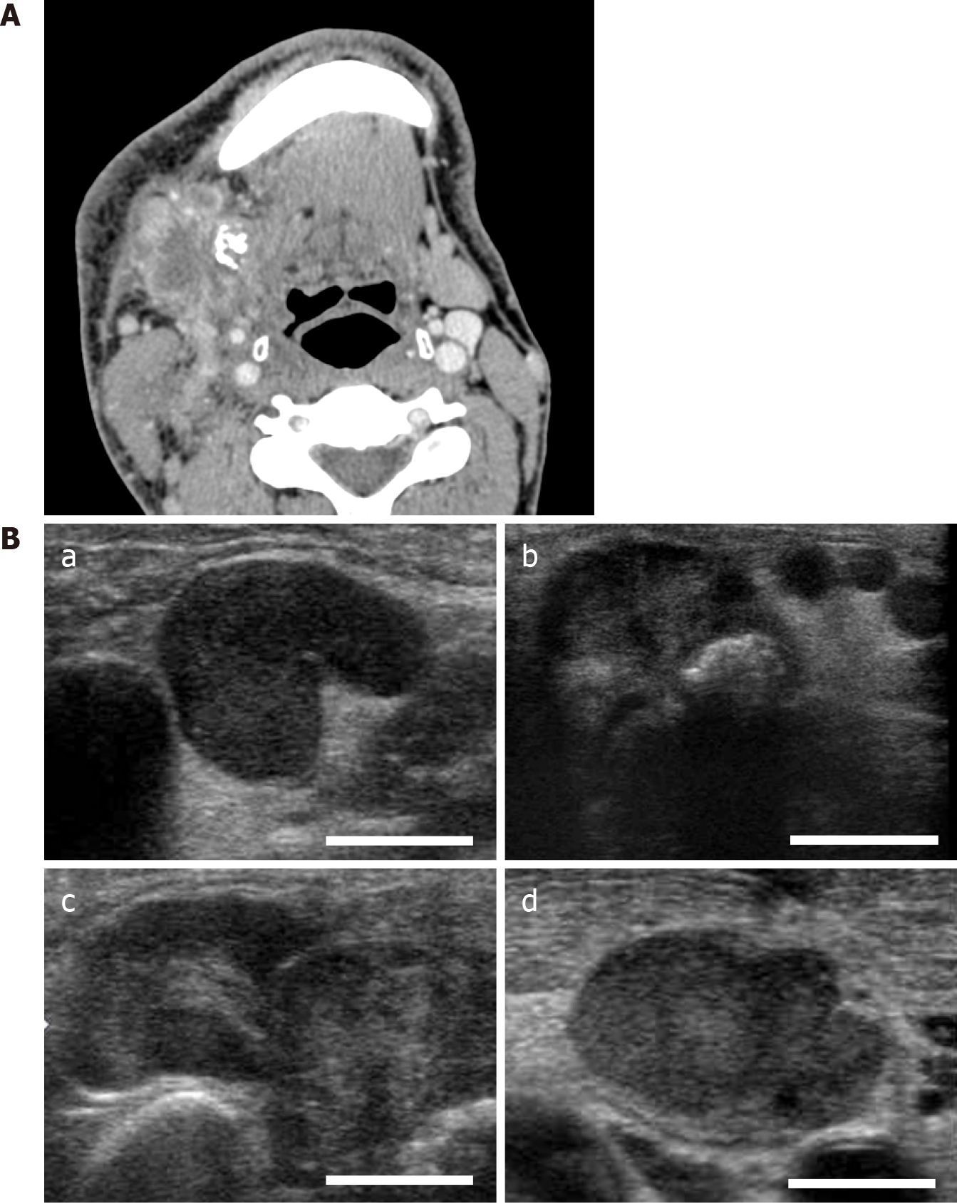
Figure 2 Contrast-enhanced computed tomographic and ultrasonographic imaging.
A: Contrast-enhanced computed tomography. Multiple cervical lymph nodes show a similar pattern, a center of low attenuation with an enhancing rim representing the central area of necrosis; furthermore, some of them displayed a tendency to fusion. A calcified body was located near these lymph nodes; B: Ultrasonography. The presence of a central echogenic hilus in the enlarged nodes keeping the oval shape and the absence of a peripheral halo (a), calcified body in the submandibular gland (b), fusion tendency of adjacent lymph nodes showing relatively strong internal echo within the mass (c), and homogeneous internal echo within the mass (d).
- Citation: Uchihashi T, Kodama S, Sugauchi A, Hiraoka S, Hirose K, Usami Y, Tanaka S, Kogo M. Salivary duct carcinoma of the submandibular gland presenting a diagnostic challenge: A case report. World J Clin Cases 2021; 9(12): 2908-2915
- URL: https://www.wjgnet.com/2307-8960/full/v9/i12/2908.htm
- DOI: https://dx.doi.org/10.12998/wjcc.v9.i12.2908


