Copyright
©The Author(s) 2021.
World J Clin Cases. Apr 26, 2021; 9(12): 2908-2915
Published online Apr 26, 2021. doi: 10.12998/wjcc.v9.i12.2908
Published online Apr 26, 2021. doi: 10.12998/wjcc.v9.i12.2908
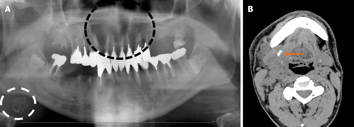
Figure 1 Radiographic imaging at the initial visit.
A: Panoramic radiographic image at the initial visit. Panoramic radiograph revealed radiolucent lesion from the left maxillary lateral incisor to the right maxillary second premolar (the black dotted line); an oval radiopaque lesion similar to sialolithiasis (the white dotted line) was also observed under the right side of the mandible; B: Non-contrast computed tomography (submandibular region). A calcified body was found near the opening of the submandibular gland (the orange arrow). Swollen cervical lymph nodes and a mass in the submandibular gland were also observed.
- Citation: Uchihashi T, Kodama S, Sugauchi A, Hiraoka S, Hirose K, Usami Y, Tanaka S, Kogo M. Salivary duct carcinoma of the submandibular gland presenting a diagnostic challenge: A case report. World J Clin Cases 2021; 9(12): 2908-2915
- URL: https://www.wjgnet.com/2307-8960/full/v9/i12/2908.htm
- DOI: https://dx.doi.org/10.12998/wjcc.v9.i12.2908


