Copyright
©The Author(s) 2021.
World J Clin Cases. Apr 26, 2021; 9(12): 2854-2861
Published online Apr 26, 2021. doi: 10.12998/wjcc.v9.i12.2854
Published online Apr 26, 2021. doi: 10.12998/wjcc.v9.i12.2854
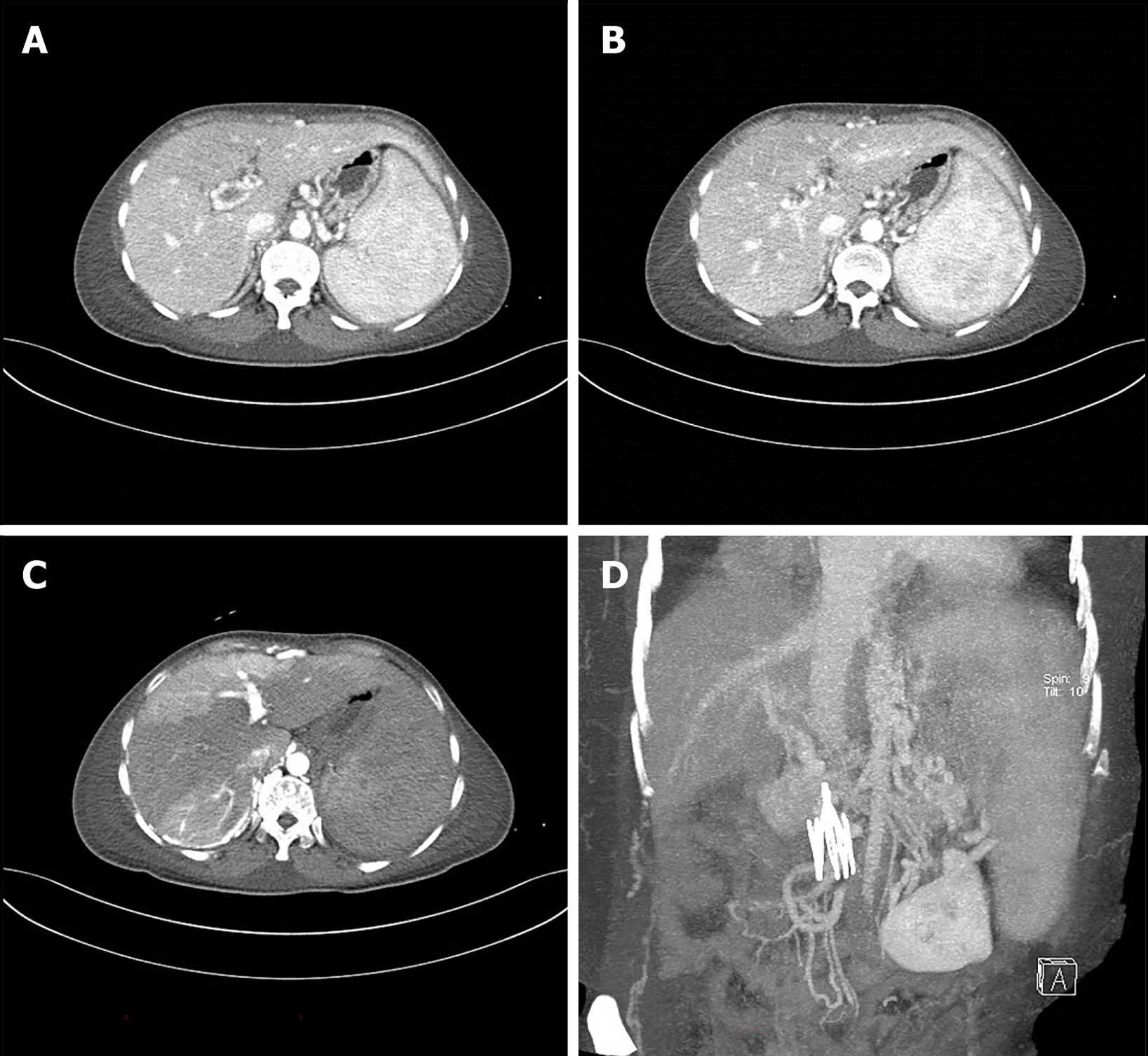
Figure 3 Abdominal vascular computed tomography.
A and B: Portal vein, splenic vein, and superior mesenteric vein embolism, collateral circulation formation, and esophageal and gastric varices. There was uneven perfusion of the liver due to portal vein thrombosis; C: The liver was clear, and the lumen was obviously narrow or occluded after implantation of the inferior vena cava filter; D: The presence of vascular filters in the lower extremities.
- Citation: Xie WX, Jiang HT, Shi GQ, Yang LN, Wang H. Behcet’s disease manifesting as esophageal variceal bleeding: A case report. World J Clin Cases 2021; 9(12): 2854-2861
- URL: https://www.wjgnet.com/2307-8960/full/v9/i12/2854.htm
- DOI: https://dx.doi.org/10.12998/wjcc.v9.i12.2854


