Copyright
©The Author(s) 2021.
World J Clin Cases. Apr 26, 2021; 9(12): 2811-2815
Published online Apr 26, 2021. doi: 10.12998/wjcc.v9.i12.2811
Published online Apr 26, 2021. doi: 10.12998/wjcc.v9.i12.2811
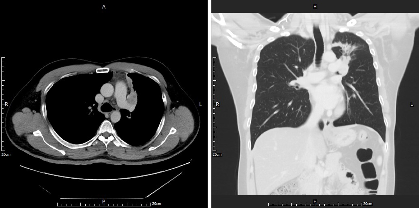
Figure 1 Chest computed tomography showed a tumor located in the left upper lobe of the lung with suspected endobronchial lesions and obstructive pneumonitis.
Vessel invasion could not be excluded.
- Citation: Yang CH, Liu NT, Huang TW. Role of positron emission tomography in primary carcinoma ex pleomorphic adenoma of the bronchus: A case report. World J Clin Cases 2021; 9(12): 2811-2815
- URL: https://www.wjgnet.com/2307-8960/full/v9/i12/2811.htm
- DOI: https://dx.doi.org/10.12998/wjcc.v9.i12.2811


