Copyright
©The Author(s) 2021.
World J Clin Cases. Apr 26, 2021; 9(12): 2791-2800
Published online Apr 26, 2021. doi: 10.12998/wjcc.v9.i12.2791
Published online Apr 26, 2021. doi: 10.12998/wjcc.v9.i12.2791
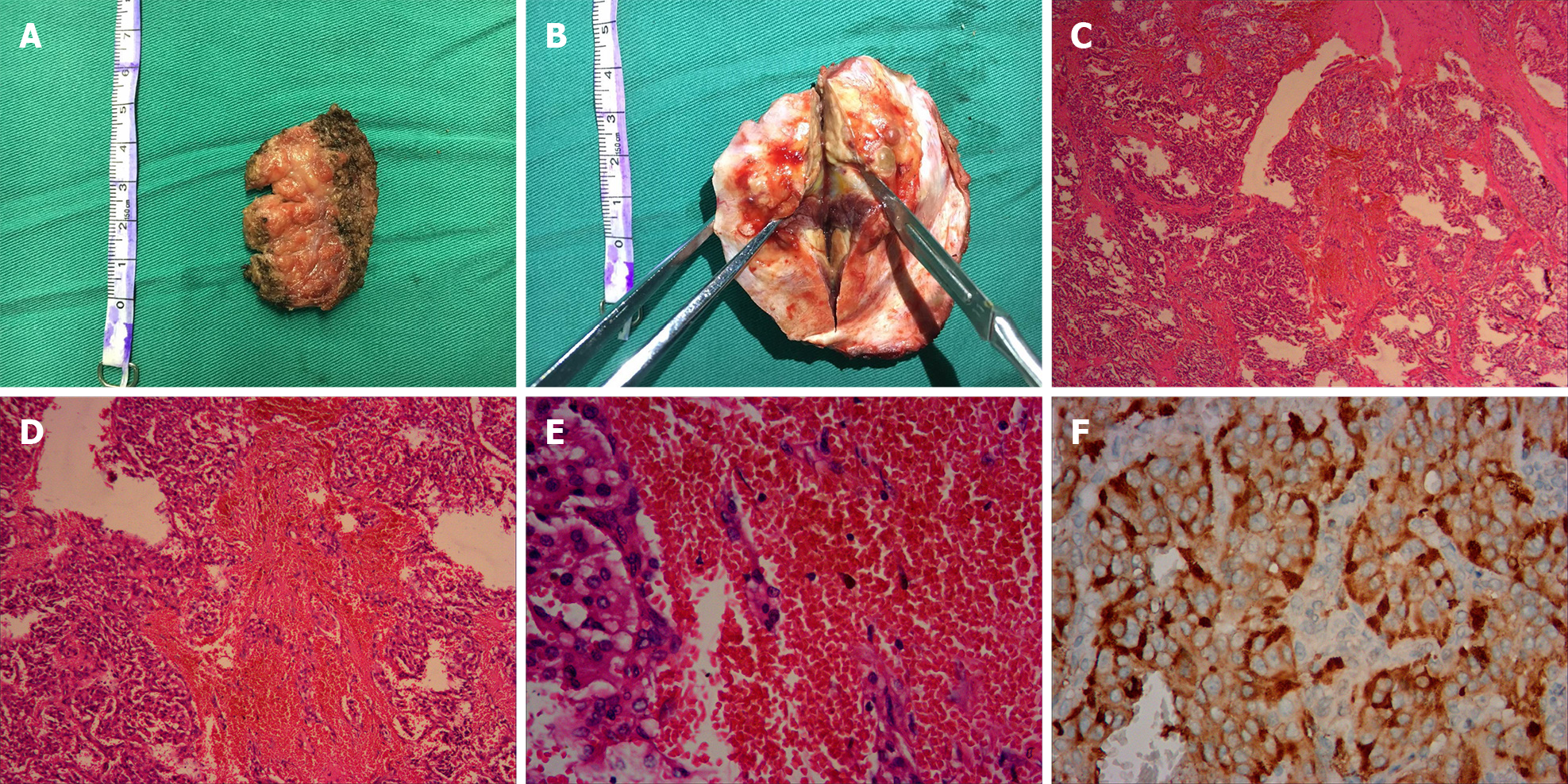
Figure 2 Histopathological appearances of the left temporal lobe mass.
A: The extracranial part (the musculi temporalis) of the gross surgical specimen; B: The intracranial part (the epidural mass with adhered temporal bone) is showed; C: Neoplastic cells arranged in a nested and trabecular fashion and surrounded by a labyrinth of capillaries are demonstrated (hematoxylin and eosin, × 40); D: Hematoxylin and eosin, × 100; E: Hematoxylin and eosin, × 400; F: Immunohistochemical analysis revealed strong diffuse immunoreactivity for CgA.
- Citation: Chen JC, Zhuang DZ, Luo C, Chen WQ. Malignant pheochromocytoma with cerebral and skull metastasis: A case report and literature review. World J Clin Cases 2021; 9(12): 2791-2800
- URL: https://www.wjgnet.com/2307-8960/full/v9/i12/2791.htm
- DOI: https://dx.doi.org/10.12998/wjcc.v9.i12.2791


