Copyright
©The Author(s) 2021.
World J Clin Cases. Apr 26, 2021; 9(12): 2739-2750
Published online Apr 26, 2021. doi: 10.12998/wjcc.v9.i12.2739
Published online Apr 26, 2021. doi: 10.12998/wjcc.v9.i12.2739
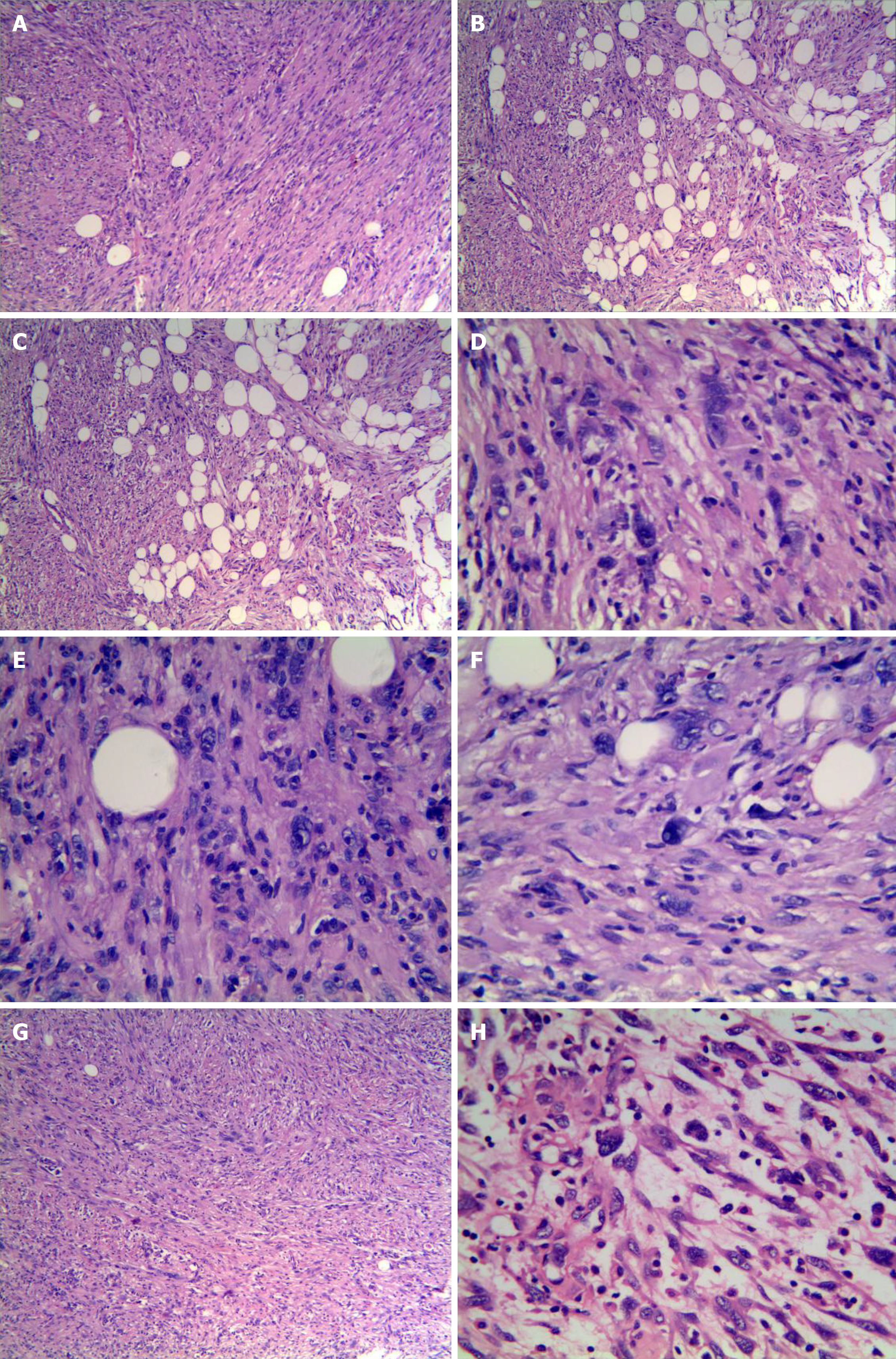
Figure 2 Histological features of the tumor stained with hematoxylin and eosin.
A: Relatively monomorphic spindled cells growing in intersecting fascicles (magnification, × 100); B: Infiltrating into the surrounding fibroadipose tissue focally (magnification, × 100); C: Epithelioid cells in the local area arranged in sheets and nests(magnification, × 400); D-F: A few scattered tumors cells exhibited irregularly hyperchromatic, bizarre, and pleomorphic nuclei with frequent intranuclear pseudoinclusions (black arrows) and rare mitotic figures(magnification, × 400); G: In some areas, tumor morphology was similar to that of inflammatory myofibroblastoma (magnification, × 100); H: In other areas, tumor morphology was similar to that of myxoinflammatory fibroblastosarcoma (magnification, × 400).
- Citation: Ding L, Xu WJ, Tao XY, Zhang L, Cai ZG. Clinicopathological features of superficial CD34-positive fibroblastic tumor. World J Clin Cases 2021; 9(12): 2739-2750
- URL: https://www.wjgnet.com/2307-8960/full/v9/i12/2739.htm
- DOI: https://dx.doi.org/10.12998/wjcc.v9.i12.2739


